Blog and News
How Does An X-ray Work?
X-ray technology was invented completely by accident. In 1895, a German physicist named Wilhelm Roentgen made the discovery while experimenting with electron beams in a gas discharge tube. Röntgen noticed that a fluorescent screen near the tube was glowing, even though the tube was insulated with cardboard. Since no light was passing through to make the screen glow, some other previously unknown form of radiation or “x-ray” must have been responsible. Röntgen noted that the ray passed through other materials as well, including human flesh! In fact, in discovering x-radiation, Röntgen also discovered its major application: imaging. The first x-ray ever produced is of Röntgen’s wife’s hand.
The point about x-rays is that, unlike visible light, they pass straight through stuff, including the stuff that people are made of. So, if you shine a beam of x-rays at someone, and put a piece of film on the other side of them, you produce a shadow of the inside of their body.
This only works at all because the different tissues that make up our bodies absorb x-rays to different extents. The problem is, though, that we consist largely of water, so most of our soft bits look much the same as far as x-rays are concerned. In fact, there are really only three tissues that are sufficiently different from each other in terms of x-ray absorption to show up on an x-ray film: bone, air and soft tissue.
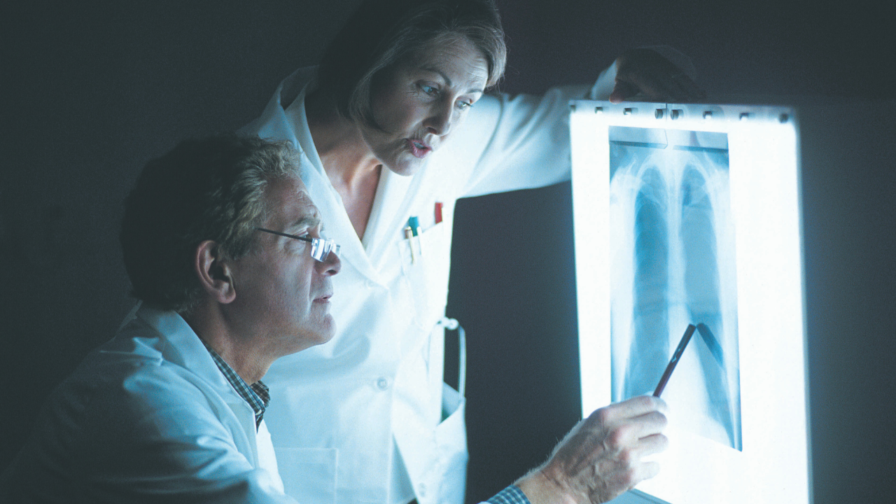
It’s the differences in those three tissues that actually make x-rays useful. In our body, soft tissue cannot absorb the high energy ray and it passes straight through. The high dense material, like bone made of calcium, do absorb the radiation. The rays that pass through to the film detector, which works much like plain camera film. The black areas are the exposed areas, representing the rays that passed through the soft tissue, while the white areas are the unexposed areas, where the rays were absorbed by tissue.
The patient is positioned between the x-ray machine and the film. It is the x ray technician’s job to properly align the patient in space in order to capture the appropriate 2-D plane. Areas not of interest should be appropriately shielded. After the beam is activated, the film is shown to the radiologist. Digital film is often used so that the image can be viewed directly on a computer monitor without true film being utilized.
And by the way, the letter “X” comes from their initial discovery, when they were an unknown form of radiation and thus given the moniker ‘”X” to indicate “unknown.”
Recent Posts


Office Hours:
Monday-Friday
8:00am-5:00pm
Saturday by appointment
Services
Contact Details
Address: 1971 Gowdey Road,
Naperville, IL 60563
Phone: 630-416-1300
Fax:
630-416-1511
Email: info@foxvalleyimaging.com
© Copyright 2023 Fox Valley Imaging, Inc..