Blog and News
Imagine you're trying to find your way through a dense fog; without a flashlight, it's nearly impossible to see what's ahead. This is akin to how radiologists feel when trying to diagnose without the use of contrast agents in X-rays. Different types of contrast agents, like barium sulfate for gastrointestinal studies or iodine-based for angiography, act as that flashlight, illuminating the path and enhancing diagnostic accuracy.
However, with each type comes its own set of challenges, risks, and benefits. You'll want to explore further how these agents can vastly improve or potentially complicate diagnostic outcomes, wouldn't you?
Key Takeaways
- Barium sulfate enhances gastrointestinal X-ray clarity, improving diagnostic accuracy.
- Iodinated compounds increase vascular structure visibility, aiding precise angiography.
- Safety profiles of contrast agents are crucial for minimizing patient risks.
- The choice of contrast agent is tailored to specific imaging needs for optimal outcomes.
Types of Contrast Agents
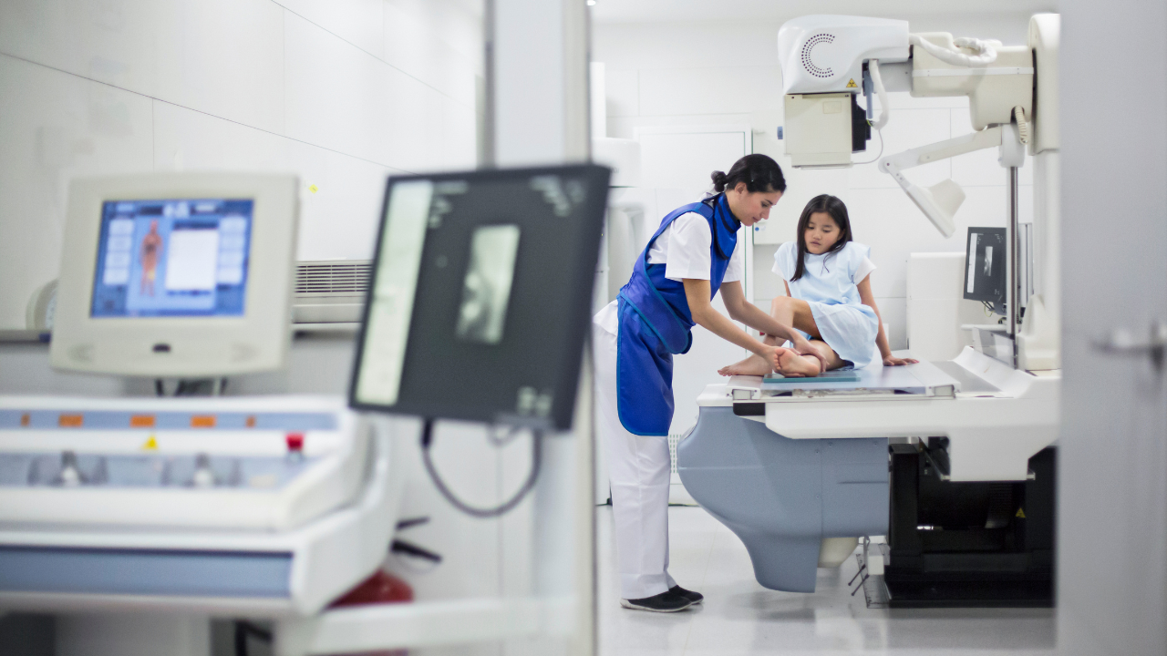
Contrast agents are essential in enhancing the visibility of internal structures during X-ray imaging. They are categorized into iodinated compounds, barium-based solutions, and gas-based agents, each tailored for specific diagnostic applications. Iodinated compounds are primarily used in vascular and organ imaging due to their high radiopacity. Gas-based agents, like air or carbon dioxide, are ideal for highlighting areas like the gastrointestinal tract, excluding barium's role.Barium Sulfate in Gastrointestinal Studies
You must consider the safety profile of barium sulfate when evaluating its use in gastrointestinal studies. Its ability to enhance image clarity greatly improves diagnostic accuracy, a key factor in medical imaging. Analyzing the balance between its safety and efficacy is vital in optimizing patient outcomes.Barium Sulfate Safety Profile
When evaluating the safety profile of barium sulfate in gastrointestinal studies, it's critical to comprehend its interactions and contraindications within the human body.- Allergic Reactions: Rare but demand immediate attention.
- Perforation Risks: Heightened in conditions like bowel obstruction or severe inflammation.
- Elimination Concerns: Confirming complete passage post-procedure to prevent impaction.
Enhancing Image Clarity
Barium sulfate greatly enhances the clarity of X-ray images in gastrointestinal studies by providing a high-contrast medium that outlines the digestive tract. Its radiopaque nature guarantees sharp demarcation between the gastrointestinal lumen and surrounding tissues, facilitating precise identification of abnormalities such as ulcers, polyps, or tumors. This diagnostic advantage enables targeted, effective treatment plans, empowering you with the freedom to pursue prime health outcomes.Iodine-Based Contrast for Angiography
Iodine-based contrast agents greatly enhance the visibility of vascular structures during angiography by selectively absorbing X-rays.- These agents reduce scatter and improve contrast resolution.
- They're tailored to highlight specific vascular territories, enabling precise diagnosis.
- Their use demands a careful balance between enhancing visibility and minimizing potential renal toxicity.
Enhancing Imaging With Contrast
You'll find that the variety of contrast agents plays a pivotal role in enhancing the clarity and accuracy of X-ray imaging. The impact these agents have on diagnostic precision can't be overstated, as they greatly improve the visibility of internal structures. Analyzing the types and their specific utility in medical diagnostics reveals the nuanced improvements in imaging outcomes.Types of Contrast Agents
Contrast agents enhance the clarity and detail of X-ray imaging by highlighting specific areas of the body, thereby improving diagnostic accuracy.- Iodine-based Compounds: Preferred for vascular and internal organ imaging due to their high atomic number, enhancing X-ray absorption.
- Barium Sulfate: Used for gastrointestinal tract visualization, offering high contrast and detailed images.
- Gadolinium: Employed in MRI scans but may be used in X-rays for soft tissue visualization.
Impact on Diagnostic Accuracy
Utilizing contrast agents greatly enhances the diagnostic accuracy of X-ray imaging by providing clearer, more detailed views of the body's internal structures. These agents differentiate between tissues, highlighting abnormalities like tumors or blockages.Risks: Allergic Reactions and Kidney Function
While enhancing diagnostic accuracy, the use of iodinated contrast agents in X-ray imaging poses risks, including allergic reactions and impaired kidney function, that require careful consideration.- Allergic responses vary, from mild hives to severe anaphylaxis.
- Contrast-induced nephropathy, potentially leading to acute kidney injury, demands attention.
- Pre-screening for allergies and evaluating renal function are critical to mitigating these risks.
Intravenous Injection Techniques
Acknowledging the risks associated with iodinated contrast agents, it's imperative to discuss the techniques of intravenous injection to minimize potential complications. Precise cannula selection, based on vein size and patient condition, is critical. Sterile technique must be maintained to prevent infection. Slow, monitored infusion allows for immediate reaction to adverse effects. These steps, rigorously followed, grant you control over the procedure, ensuring patient safety and diagnostic accuracy.Advantages of Double Contrast Studies
Double contrast studies greatly enhance the visibility of the gastrointestinal tract's interior lining, offering clearer, more detailed images for accurate diagnosis.- Improved Lesion Detection: Enables the identification of small or subtle lesions that single contrast methods might miss.
- Enhanced Mucosal Detail: Provides superior visualization of mucosal patterns, critical for diagnosing conditions like inflammation or tumors.
- Reduced Ambiguity: Minimizes interpretive errors, leading to more precise diagnostic outcomes.
Frequently Asked Questions
How Do Patient-Specific Factors, Such as Age or Pre-Existing Conditions, Influence the Choice of Contrast Agent for an X-Ray Diagnostic Procedure?
Ironically, you're not just a number. Your age and pre-existing conditions dictate the contrast agent for your X-ray, ensuring accuracy while safeguarding your health. It's freedom in healthcare, tailored to your unique story.Can the Use of Contrast Agents in X-Ray Diagnostics Interfere With Other Imaging Modalities, Such as MRI or CT Scans, if Performed in Close Succession?
Yes, contrast agents used in X-rays can affect subsequent MRI or CT scans. They may obscure details or cause artifacts, complicating interpretation. It's important to plan imaging sequences to avoid such interference.Are There Any Recent Advancements or Innovations in Contrast Agent Technology That Improve Safety or Efficacy Compared to Traditional Agents?
Yes, you're in luck—recent innovations in contrast agent technology have indeed raised the bar. They're safer and more effective, shedding light on details once in the shadows while ensuring your freedom from adverse reactions.How Does the Cost of X-Ray Diagnostic Procedures Vary With the Use of Different Types of Contrast Agents, and Does Insurance Coverage Typically Differ Among These Options?
The cost of X-ray diagnostics varies with different contrast agents, and insurance coverage often differs too. You'll find that more advanced agents may cost more but could offer better diagnostic clarity and potentially broader coverage.What Are the Environmental Impacts of Disposing of Contrast Agents Used in X-Ray Diagnostics, and Are There Any Sustainable Alternatives Currently Being Explored?
Imagine a world where your choices can protect our planet. The disposal of X-ray contrast agents poses environmental risks, yet innovative, sustainable alternatives are being explored to mitigate these impacts and preserve ecological freedom.Statistics
- The discovery of X-rays by Wilhelm Conrad Röntgen occurred in 1895, which revolutionized medical diagnostics.
- Airport security X-ray systems are designed to limit radiation exposure, with each baggage scan equivalent to flying at altitude for just two minutes due to cosmic radiation.
- A standard chest X-ray (two views) exposes a patient to about 0.1 mSv (millisieverts) of radiation, comparable to the dose received from natural background radiation over ten days.
- Approximately 6% to 8% of fractures in children are detected specifically using X-rays, underscoring their diagnostic value in pediatrics.
- Pediatric computed tomography (CT) represents only about 11% of all CT exams but may contribute as much as 35% of the total radiation dose from all medical X-rays.
- Barium studies, enhanced with contrast agents for X-ray imaging of the digestive system, account for about 8% of all diagnostic X-ray procedures.
- Dental X-rays are a significant part of dental care, with over 320 million intraoral X-rays taken annually in the United States.
- Approximately 50% of total X-ray procedures in veterinary medicine are for orthopedic examinations, aiding in the assessment of bone fractures and joint diseases in animals.
- Over 16,000 hospitals worldwide are equipped with X-ray machines, emphasizing the universal presence and reliance on this technology in healthcare facilities.
- The average number of dental X-ray films per patient per visit in the United States is 2.7, but can be higher depending on the dental condition and diagnosis protocol.
External Links
- The Health Physics Society provides insights into understanding radiation doses from X-rays and other sources. Health Physics Society - Medical X-rays FAQs
- RadiologyInfo.org offers a patient-friendly resource on how X-rays are performed and what patients can expect. RadiologyInfo.org - Chest X-ray
- The World Health Organization sections on radiation discusses the health risks and benefits related to X-rays. WHO - Radiation
- The International Atomic Energy Agency (IAEA) has resources discussing radiation protection in the use of diagnostic X-rays. IAEA - Diagnostic Radiology
- MedlinePlus, a service of the U.S. National Library of Medicine, provides a medical encyclopedia entry on X-rays. MedlinePlus - X-rays
- PhysicsCentral explores the physics behind X-rays and their applications beyond medicine. PhysicsCentral - How X-rays Work
- The British Dental Association presents guidelines for safe X-ray practices in dental care. BDA - Dental X-ray Guidelines
- The U.S. Food and Drug Administration provides guidelines on the safe use of X-rays in medical imaging. FDA - Medical X-ray Imaging
- The European Commission offers scientific guidelines on X-ray imaging and patient protection. European Commission - Radiation Protection 172
- The American Cancer Society provides a comprehensive overview of X-ray usage in mammograms for breast cancer detection. American Cancer Society - Mammograms
How to Read an X-ray Image
Reading an X-ray image requires knowledge of normal anatomy and understanding of the variations that signify disease or injury. Medical professionals are trained to examine the density and structures within an X-ray—a bright white area might indicate dense material such as bone, while darker areas indicate less dense tissues such as muscles or organs. Look for symmetry in the body structures, and note any anomalies, such as breaks in the continuity of a bone, to identify fractures. Reading an X-ray proficiently is complex and should be done by a qualified radiologist or physician.Recent Posts


Are Steroid Medications and Lifestyle Factors Triggering the Need for Bone Density Scans?
3D MRI Prostate mapping is a new procedure which offers a number of improvements compared to the traditional TRUS (transrectal ultrasound) biopsy for people who have prostate cancer. TRUS guided biopsies have been the go-to way of diagnosing prostate malignancy for the last 20 years, but this procedure is quite invasive, and it is not very precise.
Office Hours:
Monday-Friday
8:00am-5:00pm
Saturday by appointment
Services
Contact Details
Address: 1971 Gowdey Road,
Naperville, IL 60563
Phone: 630-416-1300
Fax:
630-416-1511
Email: info@foxvalleyimaging.com
© Copyright 2023 Fox Valley Imaging, Inc..