Slap Shoulder Arthrography Labral Tear
At Fox Valley Imaging we offer specialized services for the detection of labral tears.
Labrum tears often are misdiagnosed and very often surgery is scheduled for the wrong joint condition when relying on common MRI scans. Our facility is one of the few in the Chicagoland area which has the right high field imaging and specialized equipment to properly image a torn anterior or posterior labrum. If you are diagnosing a shoulder injury, considering corrective surgery, review with your surgeon the need for a better imaging to see if the labrum has been damaged.
If you are an athlete we can work with your medical team to provide the sharpest images available today along with a detailed review of any potential performance limiting issues seen in the imaging. Our Radiologists are trained in sports injury and performance.
Arthrography Tests for Shoulder Injuries
Symptoms of Shoulder Injuries
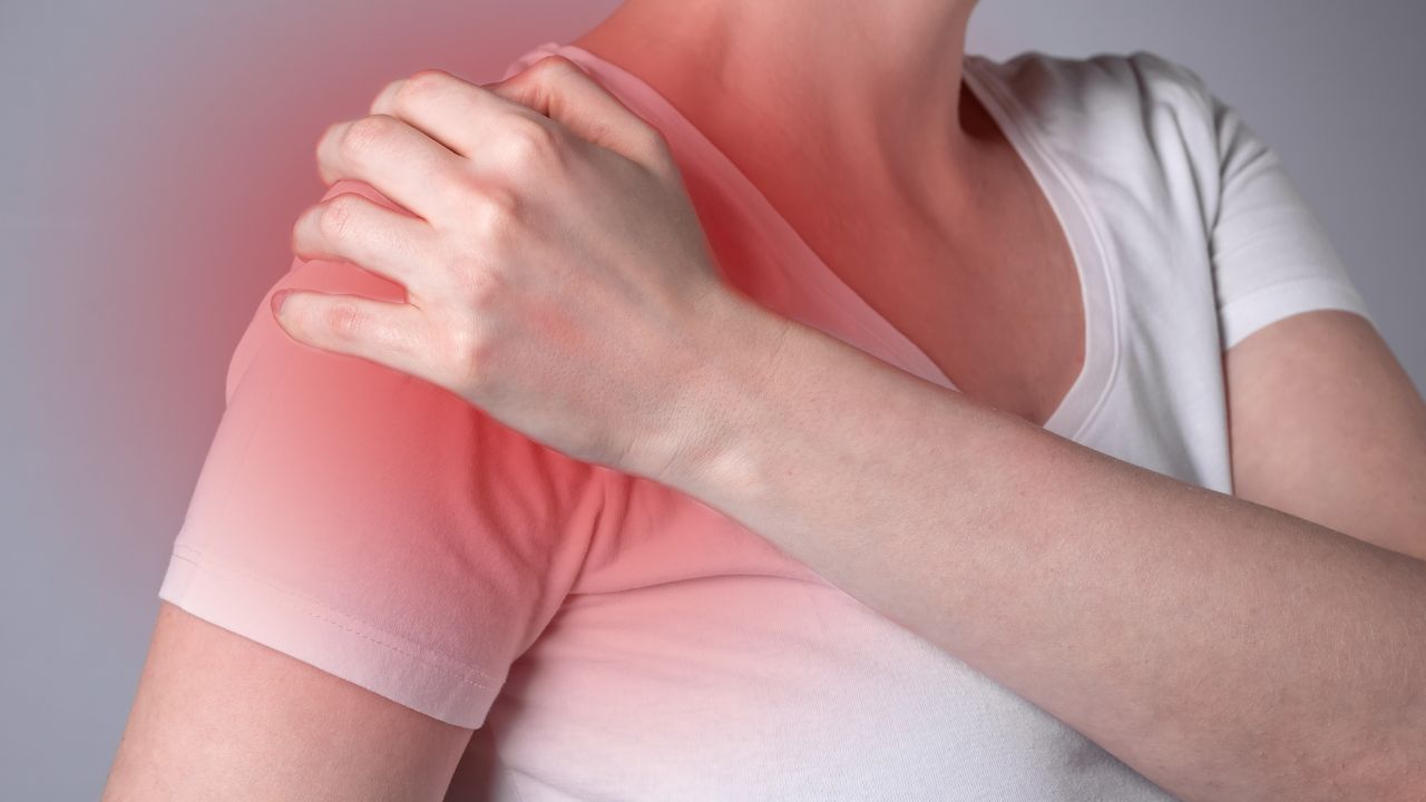
Most of the symptoms of a SLAP tear in the shoulder are very similar to those of other injuries of the shoulder. Pain, weakness, and a low motion capacity are often the most notable symptoms of the tear, which arise when performing activities that require the lifting or arms. Other notable symptoms of SLAP (Superior Labrum Anterior to Posterior) tear, at times referred to as a labral tear are locking, grinding, popping, catching, and instability in the functions of the shoulder.
Cause
The notable cause of a SLAP tear is an acute trauma to the shoulder, a good example being a fall with arms stretched out in front, sudden twist of the shoulder, falling directly on the shoulder, or a heavy pull of the arm or shoulder. The tear is common among certain athletes, especially those who engage in sports that involved repeated overhead motions, which include baseball players and bodybuilders.
What Is Tearing of the Labral?
Sometimes referred to as a SLAP tear, this is an injury of the shoulder is an injury to the shoulder cartilage in the joint called labrum, which is a ring of tissue surrounding the shoulder to cushion it from impacts. It is an important tissue that adds to the stable function of the shoulder. When torn, it may result in a partial or complete dislocation of the shoulder. The tissue is susceptible to injury especially in cases of trauma to the shoulder. The labrum will get brittle with age and can tear or fray as it ages.
How Does A Labral Tear Occur?
Labral tears happen in different forms, namely:
I. A SLAP tear – which is a superior labrum anterior and posterior tear that occurs at the area that has the biceps tendon attached to the shoulder, which is the section in the topmost part of the labrum. A SLAP tear is common in cases where a person engages in repeated high velocity overhead motions, such as athletes in sports that involve a lot of throwing. Other causes of a SLAP tear include a sudden pull or twist of the arm or shoulder, lifting heavy items, falling on the shoulder, or falling with arm outstretched.
II. A Bankart tear – which is an injury dealt to the lower section of the labrum, occurring due to the dislocation of the shoulder. The shoulder pops out of the joint tearing the labrum. Young athletes are often most susceptible to Bankart tears.
III. Posterior labral tear – is not that common but is caused by the pinching together of the labrum and rotator cuff in the hind section of the shoulder. It is a condition referred to as an internal impingement.
Symptoms of a Labral Tear
Symptoms of a labrum tear will vary depending on the location of the tear. Sometimes it can be a feeling of weakness of unstable function of the shoulder, an ache in the shoulder joint, pain when doing specific actions such as move the arm over the head, or a catching sensation when moving the shoulder among other symptoms. In some cases, though not that common, the tear might not include any pain.
How Is The Tear Diagnosed?
The deep location of the labrum tissue in the shoulder makes it somewhat difficult to make a conclusive diagnosis of the extent of the tear during a general physical exam or even based on the patients history. Proper diagnosis requires the use of an MRI and an arthrogram for the surgeon or doctor to be able to see images of the deep section of the shoulder joint. This is made possible through the injection of a contrast dye before the test. The test helps to make a proper diagnosis to determine the extent of the tear.
What Is An Arthrogram?
An arthrogram is a diagnostic exam done to check the inside of a joint- be it the wrist, shoulder, knee, or ankle – to determine the extent of injury or find the cause of a symptom a patient might claim to experience occasionally.
The first step for the test is an injection that contain a unique dye (contrast medium) that outlines the structural form of the tissue in the joint ( the cartilage and ligaments) to make them visible in form of a picture or image for medical assessment and diagnosis.
The next step is a magnetic resonance imaging, often called an MRI; this can be done without the of the contrast dye, but the use of the dye when conduction an arthrogram test (CT scan or MRI) give more accurate information for the doctor to determine where the problem lies within the joint.
Preparing For an Arthrogram?
There are no specific preparations required for an arthrogram. The general approach will include an X-ray, ultrasound, MRI, or CT scan of the shoulder joint to assess the cause of the symptom. It helps to carry any of the mentioned scans when attending an arthrogram test. The other thing to remember is to dress in something comfortable that will give an easy access to the joint during the examination.
What Happens During An Arthrogram?
The patient will lie down and the area to be examined is cleaned with an antiseptic. Next, the doctor or nurse might inject a local anesthetic to numb the area in a bit to help ease any pain that will be caused when injecting the contract dye or medium. The injection may however have a slight sting.
The patient will then have an X-ray, MRI, ultrasound, or CT scan done to aid with guidance of the insertion of the needle to the right position for the contrast die to be injected into the affect area. The injection may cause a feeling of fullness in the joint, which might have a slight pain or none at all. The type of arthrogram exam to be done and the doctor handling it will determine the type of contrast medium to be used.
For an MRI arthrogram, the doctor will include a very dilute mixture of an MRI Contrast solution called gadolinium chelates mixed with mildly salty water called sterile saline. For a CT scan arthrogram, the doctor will inject air or a little amount of X-ray contrast dye before doing the scan.
After the injections, the doctor will take the patient to respective arthrogram suite (for an MRI or CT scan) where pictures or images of the joint will be developed for medical assessment.
What Are The Effects Of An Arthrogram?
Bearing in mind that soreness in the joint is the reason why people go for the exam, most patients will experience some slight increase of the soreness a day or two after the injection and then the joint will slow return to is former state prior to having the examination done.
How Long Is The Examination?
An arthrogram exam takes around 15 minutes; however, there is a slight waiting period before the exam is done. If a CT scan will be necessary, it will take around 15 minutes while an MRI scan will take around 30 – 45 minutes. However, how long the exam takes depends on the joint being examined and the number of scans to be done. It is, therefore, better for a patient to anticipate spending around 2 hours in our facility.
Is An Arthrography Risky?
An arthrogram examination is safe and rarely has any complications.
Any adverse complication that might arise would be an infection of the joint, which might be due to the transference of an organism for the patient’s skin into the joint. As such, an arthrography is never done if the skin covering the joint is infected or broken. The risk of infections is said to be around 1 in every 40,000 people, but this is not a conclusive finding.
Cases of allergic reactions to the contrast dye injected into the joint may arise but the cases are mostly issues of a breakout of a rash, nothing very serious. Other risk such as hives and other minor infections are noted to be around 1 in every 2,000 people who take the test. Serious reactions are virtually a rare occurrence.
Cases of complication attributed to the use of the small amounts of MRI contrast dye for the arthrogram exam are yet to be reported.
The Benefits of Arthrography
The use of a contrast medium injected into the joint help to better the detection of the extent of damage to the tissue in the internal structures of the joint. Other reasons why an arthrogram exam is necessary include:
• In the shoulder: for cases where the joint is not stable or if a plain MRI or ultrasound scan showed no suspected tears of the joint tendon
• In the hip: to help identify labrum tears or condition of the joint’s rim
• In the wrist: to help identify fi the small ligaments in the wrist are torn
Bear in mind, that the doctor reserves the professional conclusion to use the additional information obtained from the arthrography test to determine the best treatment option for the patient.
Who does the Arthrogram?
In most cases, only a radiologist is to perform the arthrography test, which will include injection of the contract dye. The radiologist, who is a specialist doctor in that field, is tasked with the responsibility of ensuring the use of the right scans, injections, analyzing the scans, and developing a formal report of all the finding that will be passed to the patient’s doctor for treatment prescription.
What Is Results To Expect From An Arthrogram?
After the tests, the time it takes for the doctor to receive the formal report on the procedures will vary, based on:
• How fast the results are required
• How complex the exam was
• The necessary addition information the radiologist might request from the doctor prior to the patient having the exam
• If the patient has had prior scans done and the time to make comparisons with the finding from the new tests (which is common in situations where there is a condition or disease being followed during the assessment of the patient’s progress).
• The method use to send the report to the patient’ doctor – fax, mail, or email
Of importance:
• Be open and candid when visiting the private hospital or clinic; inquire as to when the doctor is likely to get the report.
• Discuss with the doctor about the results of the tests to make sure that ensure that you fully understand what the results mean.
Office Hours:
Monday-Friday
8:00am-5:00pm
Saturday by appointment
Services
Contact Details
Address: 1971 Gowdey Road,
Naperville, IL 60563
Phone: 630-416-1300
Fax:
630-416-1511
Email: info@foxvalleyimaging.com
© Copyright 2023 Fox Valley Imaging, Inc..