What is Echocardiography?
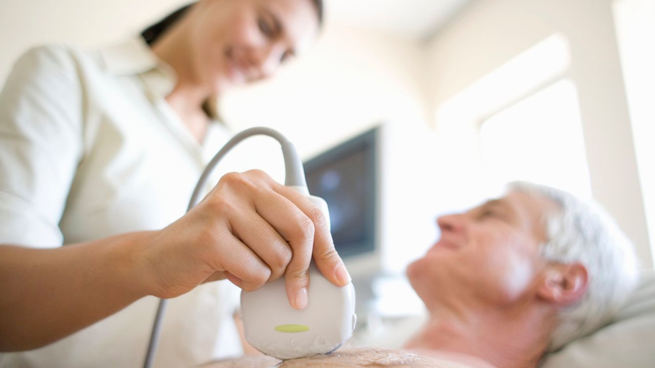
Echocardiogram, often referred to cardiac echo or simply an echo is a sonogram of the heart. Echocardiography uses standard two-dimensional, three-dimensional, and Doppler ultrasound to create images of the heart.
Echocardiography has become routinely used in the diagnosis, management, and follow-up of patients with any suspected or known heart diseases. It is one of the most widely used diagnostic tests in cardiology. It can provide a wealth of helpful information, including the size and shape of the heart (internal chamber size quantification), pumping capacity, and the location and extent of any tissue damage. An Echocardiogram can also give physicians other estimates of heart function such as a calculation of thecardiac output, ejection fraction, and diastolic function (how well the heart relaxes).
Echocardiography can help detect cardiomyopathies, such as hypertrophic cardiomyopathy, dilated cardiomyopathy, and many others. The use of Stress Echocardiography may also help determine whether any chest pain or associated symptoms are related to heart disease. The biggest advantage to echocardiography is that it is noninvasive (doesn’t involve breaking the skin or entering body cavities) and has no known risks or side effects.
Not only can an echocardiogram create ultrasound images of heart structures, but it can also produce accurate assessment of the blood flowing through the heart, using pulsed or continuous wave Doppler ultrasound. This allows assessment of both normal and abnormal blood flow through the heart. Color Doppler as well as spectral Doppler is used to visualize any abnormal communications between the left and right side of the heart, any leaking of blood through the valves (valvular regurgitation), and to estimate how well the valves open (or do not open in the case of valvular stenosis).
Preparing for an Echocardiogram
Content coming soon