Blog and News
Computed tomography (CT) significantly enhances your cancer management strategy. By providing high-resolution images, CT scans allow precise visualization of tumors, showing their size, shape, and exact location within your body. This level of detail aids in accurately staging your cancer, crucial for tailoring your treatment effectively. CT scans also guide biopsy procedures, ensuring accuracy while minimizing risks. Throughout your treatment, they monitor responses and adjust plans as needed, detecting any changes in the tumor or potential spreading to other areas. This tool is vital in tracking treatment effectiveness
and adapting strategies to optimize your care outcomes. Discover how this technology could specifically benefit your treatment plan.
Key Takeaways
- CT scans provide detailed images, aiding in accurate tumor visualization, staging, and monitoring treatment response.
- Helps guide biopsy procedures, ensuring precise needle placement and safer tissue sampling.
- Enables the detection of metastasis by identifying the spread of cancer to other body parts.
- Assists in lymph node analysis, crucial for detecting early metastasis and influencing treatment decisions.
- Follow-up CT scans are vital for assessing the effectiveness of treatment and detecting any recurrence.
Tumor Visualization Techniques
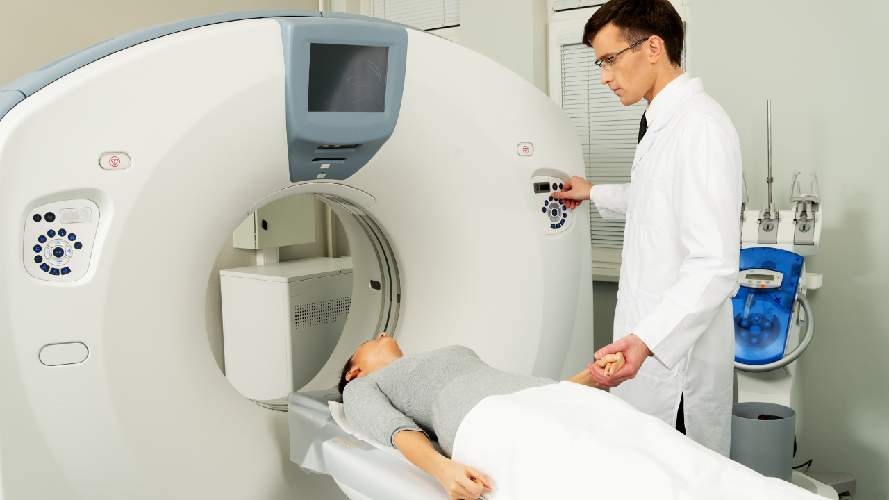
Computed tomography (CT) excels in mapping out tumors by providing high-resolution images that highlight the size, shape, and location of cancerous growths within the body. When you're tasked with diagnosing or monitoring a patient's cancer progression, the clarity and detail offered by CT scans are indispensable. They allow you to discern even subtle differences in tissue density, facilitating a more precise analysis of tumor boundaries and heterogeneity. CT scans employ X-rays in a finely tuned manner, rotating around the patient to capture multiple cross-sectional images from various angles. These images are then reconstructed digitally to form a comprehensive 3D model of the tumor and surrounding tissues. This process, known as tomographic imaging, is crucial for understanding the complex anatomy and the exact extent of the tumor without invasive procedures. Moreover, the speed at which CT scans are conducted minimizes patient discomfort and reduces the risk of movement artifacts in images. This rapid imaging capability is essential, especially in emergency situations where swift diagnosis can significantly alter treatment decisions. As you continue to use CT technology, remember that your ability to interpret these detailed images directly impacts your effectiveness in aiding those battling cancer.Staging Cancer Accurately
Accurately staging cancer is crucial for determining the most effective treatment plan and predicting patient outcomes. Computed Tomography (CT) plays a pivotal role in this process by providing detailed cross-sectional images of the body. These images enable oncologists to assess the size, shape, and location of tumors with precision. You'll find that CT scans are integral in identifying the extent to which a cancer has spread or metastasized. This is particularly vital for solid tumors located in areas like the lungs, liver, and pancreas. By determining the stage of cancer, doctors can tailor treatment approaches, decide whether surgery is a viable option, and predict possible recovery scenarios. For instance, a Stage I lung cancer, localized and small, might be managed differently compared to a Stage IV lung cancer, which has spread to distant organs. Moreover, the ability of CT to monitor tumor response to therapy over time helps in adjusting treatment plans as needed, ensuring that you're providing patient-centered care. This adaptability not only maximizes the likelihood of successful outcomes but also contributes to a more informed and compassionate approach to cancer treatment. By leveraging this technology, you're equipped to offer hope and precise care to those you serve.Guiding Biopsy Procedures
When you use computed tomography (CT) to guide biopsy procedures, you enhance the precision of selecting the biopsy site, crucial for accurate diagnosis. This approach significantly reduces the risks of complications, as it allows for real-time visualization and precise needle placement. You'll find that integrating CT in biopsy procedures supports a safer, more efficient pathway to obtain critical tissue samples for cancer analysis.Biopsy Site Precision
Guiding biopsy procedures with precision, computed tomography (CT) enables doctors to pinpoint the exact location of a tumor for tissue sampling. This accuracy is crucial, as the exact positioning of the biopsy needle can significantly influence the quality of the sample obtained, which in turn affects the diagnostic accuracy. By using CT images, you're not just guessing where to place the needle; you're using a detailed, real-time map of the patient's internal anatomy. This approach reduces the likelihood of multiple needle insertions, making the process more efficient and patient-friendly. Furthermore, it ensures that the sample is representative of the tumor, providing reliable data for pathology. In essence, CT-guided biopsies are a cornerstone in devising effective treatment plans for cancer patients.Reducing Complication Risks
Utilizing computed tomography during biopsy procedures significantly reduces the risk of complications by ensuring the needle's precise placement in tumor tissue. As you guide the needle, CT imaging provides real-time, high-resolution images that help distinguish between different types of tissues, ensuring the target area is accurately reached without damaging surrounding structures. This precision is particularly crucial in densely packed anatomical regions where traditional methods risk puncturing vital organs or blood vessels. Moreover, the detailed visualization afforded by CT scans allows for the collection of adequate tissue samples, which are essential for accurate diagnosis and treatment planning. By minimizing the number of attempts needed to retrieve a viable sample, you also reduce the patient's exposure to potential pain and infection, enhancing overall procedural safety and efficacy.Monitoring Treatment Response
Computed tomography (CT) scans are crucial for assessing how effectively your cancer treatment is working, providing detailed images that reveal changes in tumor size and structure. As you undergo therapy, these scans become a key tool in determining whether the cancer cells are responding to the treatment. They allow your healthcare team to visualize reductions in tumor mass and modifications in its density, offering insights that are vital for tailoring your treatment plan. CT imaging is adept at capturing cross-sectional views of your body, which can be meticulously analyzed to detect even subtle changes in the tumor's response to chemotherapy, radiation, or other therapeutic interventions. This capability is essential for oncologists to make informed decisions about continuing, adjusting, or changing your treatment regimen. It's not just about tracking the shrinkage of tumors; CT scans also help in assessing the effectiveness of the treatment on affected lymph nodes and surrounding tissues, ensuring that the entire area of concern is responding as expected.Identifying Metastasis
CT scans play a crucial role in detecting the spread of cancer to other parts of your body, known as metastasis. This imaging technology is pivotal in both the early detection and ongoing management of metastatic cancer, helping healthcare providers to devise and refine treatment plans that are as effective as possible. Here's how CT imaging stands out in identifying metastasis:- High-Resolution Images: CT scans provide detailed cross-sectional images of your body, allowing oncologists to view small nodules or tumors that might be metastases. This capacity for detailed visualization is crucial for early detection, which can significantly influence treatment decisions and outcomes.
- Whole-Body Scanning Capability: Unlike some other imaging modalities that only capture specific areas, CT scans can cover practically the whole body in a single session. This is essential for checking if cancer cells have spread to distant organs, providing a comprehensive overview of your health status.
- Speed and Accuracy: CT scans aren't only detailed but also fast. They can quickly scan large areas, which is vital for timely diagnosis and treatment planning. This speed doesn't compromise the accuracy, making CT an invaluable tool in the fight against cancer.
Analyzing Lymph Nodes
You'll find that computed tomography (CT) is instrumental in lymph node mapping, providing high-resolution images that detail the size and location of nodes. This precision is crucial for identifying metastatic spread early, which significantly impacts treatment options and outcomes.Lymph Node Mapping Precision
Accurately mapping lymph nodes using computed tomography significantly enhances the precision of cancer staging and targeted treatment strategies. By enabling clinicians to visualize the size and location of lymph nodes, CT scans are indispensable in determining the extent of cancer and planning the optimal course of treatment. Here's how CT imaging impacts lymph node analysis:- Size and Shape Assessment: CT scans provide detailed images that reveal the dimensions and morphology of lymph nodes, crucial for assessing potential abnormalities.
- Location Accuracy: It pinpoints the exact anatomical locations of lymph nodes, vital for surgical planning and radiation therapy.
- Consistency and Repeatability: CT offers a reliable means to monitor lymph node changes over time, supporting ongoing assessment and adjustments to treatment plans.
Identifying Metastatic Spread
Through the analysis of lymph nodes, computed tomography (CT) plays a pivotal role in identifying metastatic spread, essential for crafting precise cancer treatment protocols. As you delve into CT scans, you're not just looking at images; you're examining potential sites of cancer spread which are critical for staging the disease and planning the pathway of intervention. CT's high-resolution capabilities allow you to discern even subtle changes in the lymph nodes, which might indicate metastases. This accuracy is vital because lymphatic spread can be the first sign of cancer dissemination beyond the primary site.Expertise of Radiologists
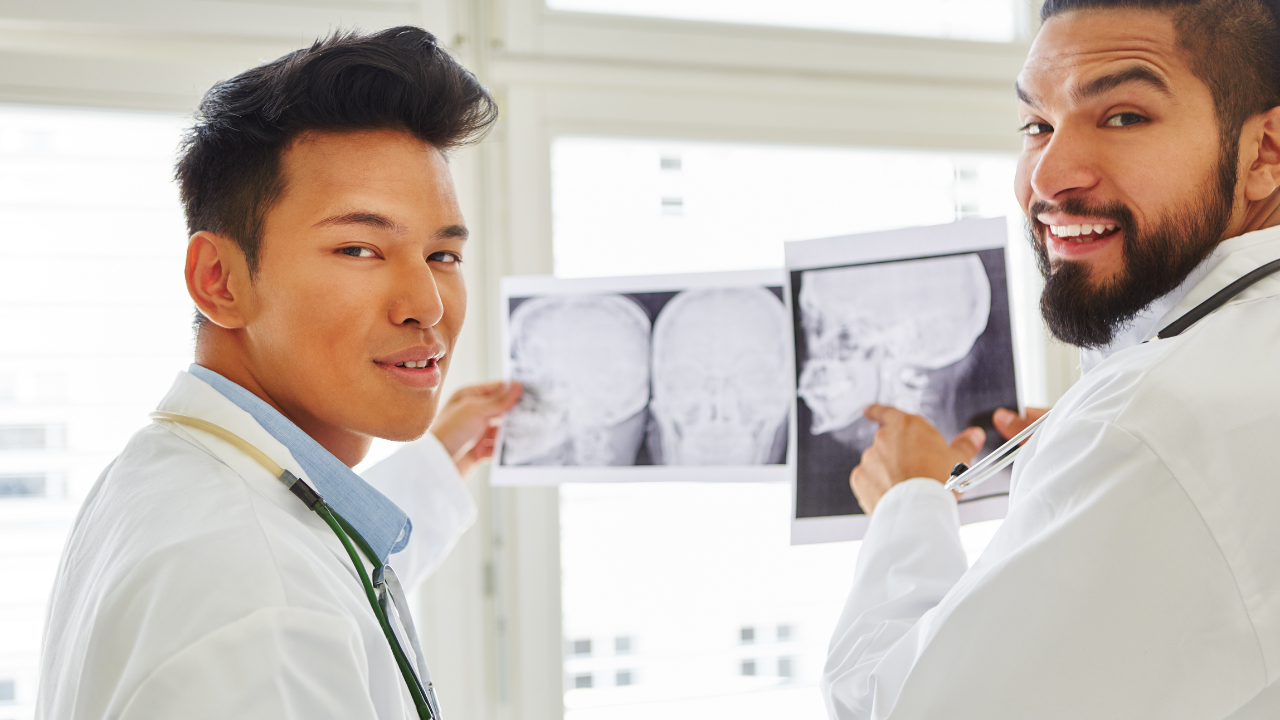
Radiologists play a crucial role in interpreting CT scans to ensure precise diagnosis and effective treatment planning for cancer patients. Their expertise is pivotal in distinguishing between benign and malignant lesions, assessing the extent of the disease, and aiding in the decision-making process for treatment strategies. Here are three key aspects of their expertise:- Advanced Training: Radiologists undergo extensive training in imaging interpretation, including mastery of various imaging modalities and understanding of complex anatomical and pathological conditions associated with cancer. They're adept at recognizing subtle changes in images that might indicate early stages of cancer.
- Interdisciplinary Collaboration: They work closely with oncologists and surgeons, providing critical insights that influence treatment plans. Their ability to interpret imaging results accurately ensures that all members of the cancer care team are well-informed, facilitating a coordinated approach to patient management.
- Continual Learning: The field of radiology is ever-evolving with advancements in technology and imaging techniques. Radiologists must stay abreast of these developments to provide the best care. They frequently participate in research and continuous education to refine their skills and update their knowledge base.
Conducting Follow-Up Scans
Follow-up CT scans are essential for monitoring the effectiveness of cancer treatment and detecting any recurrence of the disease. As you guide patients through their treatment journey, understanding the role of these scans can empower you to provide better care. After the initial treatment, whether it's surgery, chemotherapy, or radiation therapy, CT scans are routinely employed to assess the state of the disease. The timing of follow-up scans varies based on the type, location, and stage of cancer, as well as the specific treatment protocols used. Typically, the first scan is conducted a few months after treatment completion to establish a baseline post-treatment state. Subsequent scans are then scheduled at intervals that your oncological team finds most appropriate, often every 3 to 6 months for the first couple of years. During follow-up, attention isn't just on detecting recurrence; you're also looking for possible metastasis or secondary cancers, which are critical for timely intervention. Moreover, these scans help in evaluating the patient's response to therapy, informing potential adjustments in the treatment plan. It's crucial to keep your patients informed and prepared for each step, reducing anxiety and fostering a collaborative environment in their care pathway.Precision in Imaging
Precision in imaging plays a crucial role in enhancing the accuracy of CT scans used in cancer diagnosis and treatment monitoring. As you're dedicated to serving patients effectively, understanding the technical nuances of CT imaging is pivotal. Here are three key aspects where precision matters:- Detection of Small Tumors: High-resolution CT imaging allows for the detection of minute tumors that might be overlooked in less precise scans. Early detection is crucial for successful treatment outcomes and can significantly alter a patient's treatment pathway.
- Tumor Characterization: Precision in imaging provides detailed insights into the tumor's morphology and composition. This information is vital for oncologists to determine the nature of the cancer, whether it's aggressive, and how it's likely to respond to different treatments.
- Monitoring Treatment Response: Accurate and precise imaging is fundamental in assessing how well a cancer is responding to treatment. Changes in tumor size, density, or metabolic activity, which are captured through enhanced imaging techniques, guide oncologists in tweaking or continuing treatment protocols.
10 Essential Insights into Computed Tomography: Understanding CT Scans Inside Out
Functional CT Scanning: Perfusion Studies
Beyond anatomical imaging, CT technology extends to functional studies, such as perfusion CT, which measures blood flow to various tissues. These studies are particularly significant in stroke management, where they help determine the affected brain areas and the extent of the blood supply compromise, thereby informing acute treatment decisions and subsequently improving patient outcomes.Choosing Between CT and MRI
CT scans and MRI scans both offer detailed images of the body's internal structures but use different technologies and are chosen based on the patient's condition. CT scans are preferred for their speed and efficacy in emergency situations, as well as for visualizing bone fractures and chest imaging. MRI, on the other hand, is chosen for its superior soft tissue contrast and the fact that it doesn't use ionizing radiation, making it suitable for detailed joint and brain imaging.Preparation Processes for Patients Undergoing CT
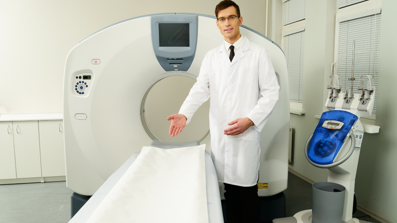
Prior to a CT scan, patients may be advised to abstain from eating or drinking for several hours, particularly if a contrast agent will be used. Clothing with metal fasteners and jewelry should be removed to prevent interference with the imaging. The patient will be positioned on a motorized table which slides into the CT scanner, and they will need to stay motionless during the scan to ensure clear images are captured.Pediatric Considerations for Computed Tomography
When using CT scans for children, special considerations are taken to minimize radiation exposure, adhering to pediatric protocols that adjust the radiation dose based on the child's size. As children are more sensitive to radiation, healthcare providers practice judicious use of CT and opt for alternative imaging methods like ultrasound or MRI when possible.Post-Processing Techniques in CT Imaging
After CT data is acquired, post-processing techniques are used to refine images for clearer interpretation. Techniques like multiplanar reconstruction (MPR), volume rendering, and maximum intensity projection (MIP) assist radiologists in analyzing data more effectively. These tools enhance diagnostic accuracy by providing different perspectives and detailed views of the scanned tissues.Applications of CT in Dental and Maxillofacial Surgery
CT scans have revolutionized dental and maxillofacial treatments by enabling precise 3D imaging of the teeth, jaw, and facial bones. They assist in the assessment for dental implants, reveal complex tooth root structures, and help diagnose temporomandibular joint (TMJ) disorders, among other applications. The clear images are integral in surgical planning and ensuring successful outcomes in dental procedures.The Role of CT in Cancer Detection
CT scans play a central role in the early detection and management of cancer. They provide detailed imagery that helps in locating tumors, understanding their size and shape, and determining if the disease has spread. These scans are also integral in planning cancer treatment such as surgery or radiation therapy, and in monitoring how the cancer responds to treatment over time.Understanding CT Scan Safety Measures
While CT scans are routine and generally safe, they do involve exposure to ionizing radiation. The medical community takes this seriously, and as such, numerous safety protocols are in place. These include using the lowest possible radiation dose, shielding to protect nearby body parts, and limiting the frequency of scans. Healthcare providers are committed to patient safety, following the principle of ALARA (As Low As Reasonably Achievable) when it comes to radiation exposure.Advancements in CT Imaging: 3D Reconstruction
One of the most significant advancements in CT imaging is the development of 3D reconstruction techniques. These reconstructions enable clinicians to visualize complex anatomical structures in three dimensions, offering a more comprehensive understanding of the patient's anatomy and pathology, which is particularly helpful in surgical planning and in educational settings for patient and professional understanding.Contrast Agents in Computed Tomography
To enhance the clarity of CT images, contrast agents are sometimes used. These substances, which can be ingested or injected, help to highlight specific areas of the body by increasing the contrast on the images. As a result, they aid in the better differentiation of organs, blood vessels, and potential abnormalities. However, their use requires careful patient screening for allergies and kidney function to prevent adverse reactions.Frequently Asked Questions
What Are the Risks Associated With CT Scans for Cancer Patients?
- CT scans expose you to radiation, which can slightly increase cancer risk. It's crucial to balance this risk against the scan's diagnostic benefits, ensuring it's justified by your medical needs and situation.
How Long Does a Typical CT Scan Take?
- A typical CT scan takes about 10 to 30 minutes, depending on the area being examined. It's relatively quick, allowing you to get back to your day with minimal disruption.
Can Children Undergo CT Scans for Cancer Detection?
- Yes, children can undergo CT scans for cancer detection. These scans are tailored with lower doses to minimize radiation exposure, ensuring the procedure is both safe and effective for younger patients.
Are There Alternatives to CT Scans for Cancer Diagnosis?
- Yes, there are alternatives to CT scans for diagnosing cancer. You might consider MRI, ultrasound, or PET scans, which offer different benefits and can be less invasive or expose you to less radiation.
How Often Should Cancer Patients Get CT Scans?
- Opening Pandora's box, the frequency of CT scans for cancer patients varies. It depends on the type, stage of cancer, and treatment response. Consult with your oncologist to tailor a schedule that fits your needs.