Blog and News

To effectively train students in heart imaging techniques, you'll want to integrate the latest research and develop comprehensive outlines. Start by familiarizing them with probe handling, ensuring they understand the probe's orientation and settings. Practice image acquisition is crucial; position patients properly and adjust the probe to capture clear images. Review anatomical landmarks extensively, identifying key structures like heart chambers and valves. Implement measurement protocols for precision and foster a robust feedback system to refine techniques. By encouraging questions and using virtual reality tools, you maintain an engaging learning environment. Following these steps, your students will gain a deeper insight into sophisticated imaging strategies.
Listen to the Article
Key Takeaways
- Utilize interactive and virtual reality tools to simulate real-life echocardiography scenarios for hands-on practice.
- Incorporate regular feedback sessions to guide improvements in probe handling and image interpretation.
- Conduct thorough reviews of anatomical landmarks and measurement techniques to ensure accuracy in diagnoses.
- Foster an environment that encourages questions and critical thinking to deepen understanding of echocardiographic principles.
- Assess student progress through both formative and summative evaluations to monitor and enhance learning outcomes.
Preparing Teaching Materials
To effectively prepare teaching materials for heart imaging techniques, begin by gathering the latest research and clinical guidelines relevant to the topic. You'll want to ensure that the information you collect is up-to-date and reflects the current standards of care. This involves reviewing recent publications in cardiology journals, guidelines from heart associations, and updates from health conferences.
Once you've accumulated the necessary data, distill it into key concepts that are essential for understanding heart imaging. Create outlines that cover topics like the types of heart imaging modalities, the anatomy relevant to each technique, and the specific indications for their use. Remember, your goal is to make complex information accessible and digestible for your students.
Next, develop visual aids. High-quality images and diagrams are crucial as they help illustrate structures, functions, and procedures. Consider incorporating interactive elements such as videos and animations that demonstrate dynamic processes like blood flow and heart valve functions.
Demonstrating Probe Handling
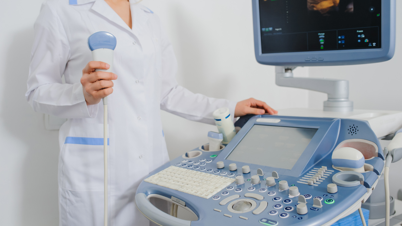
Before you start using the imaging probe, familiarize yourself with its handling and adjustments to ensure precise imaging results. Hold the probe firmly yet gently, as you'd a delicate instrument. Your grip should be comfortable, allowing for subtle movements without strain. Notice the probe's orientation marker; this small notch or line indicates the direction of the imaging plane and is crucial for accurate placement.
As you hold the probe, practice maneuvering it with slight rotations of your wrist. This skill is key in adjusting the probe's angle and position to capture optimal images of the heart's structures without needing to reposition the entire device frequently. Remember, your adjustments should be smooth and controlled. Jerky movements can lead to unclear images and may discomfort the patient.
Ensure you're also familiar with the probe's buttons and settings. These control the depth and focus of the imaging, which are essential for tailoring the echocardiogram to each patient's specific needs. Mastering these adjustments before you begin actual scanning not only saves time but also enhances the comfort and safety of the patient, aligning with your dedication to serving others through high-quality medical care.
Practicing Image Acquisition
Having mastered the basics of probe handling, you're now ready to practice image acquisition to capture clear and effective heart images. First, ensure you've set up the echocardiogram machine correctly. Check the settings for brightness and contrast to suit your specific examination requirements. You'll want to adjust these settings depending on the patient's body type and the depth of the heart within the chest cavity.
Next, position the patient properly. They should lie on their left side, which brings the heart closer to the chest wall, offering better visualization. Make sure they're comfortable, as patient movement can disrupt the imaging process.
Now, focus on obtaining a stable hand position. A steady hand reduces blurring and helps in capturing high-quality images. Practice moving the probe slowly and deliberately, maintaining slight but consistent pressure against the chest. This technique reduces the amount of air between the probe and the skin, which can interfere with image clarity.
You'll also need to continuously adjust the angle of the probe to explore different views of the heart. Each movement should be smooth and controlled, aiming to cover all necessary angles without rushing, ensuring each image you capture is as informative as possible.
Reviewing Anatomical Landmarks
Once you've captured initial heart images, it's crucial to review anatomical landmarks to ensure accuracy and completeness in your assessment. You'll need to identify key structures such as the chambers- the left and right atria and the ventricles. Pay close attention to the valves: the tricuspid, pulmonary, mitral, and aortic valves. Each of these landmarks plays a critical role in diagnosing and monitoring heart health, and your ability to accurately locate and identify them can make a significant difference in patient outcomes.
Start by locating the mitral valve, easily identifiable as it lies between the left atrium and left ventricle. This is a common site of various cardiac pathologies and understanding its position and appearance is essential. Progress to the aortic valve, situated at the base of the heart, leading from the left ventricle to the aorta. Remember, the orientation and symmetry of these valves are as important as their appearance.
Ensure you're also familiar with the septum, the dividing wall between the left and right sides of the heart. A clear view of the septum can help you assess its integrity and thickness, crucial for diagnosing conditions like hypertrophy or defects.
Instructing on Measurement Techniques
To ensure consistent results in heart imaging, you'll need to standardize measurement protocols across all examinations. Start by calibrating your equipment accurately; this step is crucial for enhancing your skills in measuring heart structures precisely.
Focus on repetitive practice to improve your accuracy, making sure each measurement adheres to the established guidelines.
Standardizing Measurement Protocols
First, ensure you're familiar with the precise steps involved in using the standardized measurement protocols for heart imaging. You'll need to consistently apply these protocols to every examination to ensure accuracy and reliability. Start by calibrating the echocardiography machine according to the manufacturer's instructions. This is crucial for maintaining the integrity of the measurements you'll be taking.
Next, systematically follow the protocol for each type of measurement. For instance, when measuring the left ventricular dimensions, position the calipers exactly as stipulated in the guidelines. It's essential to avoid any deviation that could lead to variability in your results. Remember, your adherence to these standardized protocols not only supports your development as a competent technician but also ensures you provide the best possible care to your patients.
Enhancing Accuracy Skills
Mastering precise measurement techniques is crucial for enhancing your accuracy skills in heart imaging.
First, familiarize yourself with the standard measurement guidelines established for echocardiography. You'll need to consistently apply these guidelines to ensure accurate and reliable results.
Begin by practicing on phantom models or simulations that provide opportunities to refine your technique without risk. Pay close attention to calibrating your equipment correctly; even minor discrepancies can lead to significant errors in measurement.
When measuring, use a steady hand and avoid rush. Detail and patience are your allies here. Regularly participate in peer reviews and feedback sessions to learn from others' insights and improve your precision.
Simulating Clinical Scenarios
As you move into simulating clinical scenarios, you'll begin by role-playing patient cases to enhance your diagnostic acumen.
You'll also incorporate virtual reality tools, which provide a highly immersive environment for practicing complex heart imaging procedures.
This approach ensures you're not just learning passively but actively engaging with realistic clinical challenges.
Role-Playing Patient Cases
To enhance your understanding of heart imaging techniques, engage in role-playing patient cases to simulate real-life clinical scenarios. You'll tackle a variety of patient profiles, each presenting unique challenges and learning opportunities. Begin by reviewing the patient's history and symptoms as presented in your case study. You'll then decide which imaging techniques are most appropriate for diagnosing the patient's heart condition.
As you proceed, interpret the imaging results with precision, considering how the findings correlate with the patient's symptoms and history. Discuss your diagnostic reasoning with peers or instructors, which fosters a collaborative learning environment and enhances critical thinking skills. This hands-on approach not only prepares you for actual clinical situations but also ingrains a compassionate understanding of patient care in your practice.
Utilizing Virtual Reality Tools
Building on role-playing patient cases, you can further enhance your skills by utilizing virtual reality tools to simulate complex clinical scenarios in heart imaging. These tools allow you to immerse yourself in highly realistic, interactive environments where you can practice and refine your echocardiography techniques without the risk associated with real-life procedures.
Start by selecting a scenario that matches the specific skills you're aiming to improve. You'll navigate through virtual patient interactions, making real-time decisions based on the imaging data you collect. Pay close attention to the feedback provided by the simulation; it's crucial for identifying areas where you need improvement.
Providing Feedback on Technique
When providing feedback on heart imaging techniques, it's crucial to focus on specific areas for improvement, ensuring the feedback is both constructive and actionable. As an educator, you'll want to observe the student's handling of the ultrasound probe, their ability to position it correctly, and the clarity with which they capture images. If a student misaligns the probe, don't just note the error; demonstrate the correct angle and explain how proper alignment enhances image quality and diagnostic accuracy.
Next, assess their interpretation of the images. If you notice inaccuracies, pinpoint the specific areas where the errors occurred. Offer clear, step-by-step guidance on how to identify key structures and boundaries in the heart. This might involve showing them examples of both poor and optimal images, discussing the differences, and suggesting strategies to improve their observational skills.
Encouraging Questions
As you progress in your heart imaging training, it's essential to foster curiosity by encouraging questions. This approach not only clarifies your understanding but also deepens your engagement with the material.
Fostering Curiosity During Lessons
Why not start by encouraging your students to ask questions throughout the lesson, thus sparking their curiosity and enhancing their understanding of heart imaging techniques? As you introduce new concepts, pause frequently and invite your students to probe deeper into the subject matter. This interaction not only clarifies their doubts but also helps them connect theoretical knowledge with practical application.
Make it a habit to create an open, inclusive atmosphere where students feel comfortable voicing their thoughts without fear of judgment. Use probing questions like 'What do you think happens if...?' to stimulate further discussion and exploration.
Benefits of Inquiry-Based Learning
Encouraging questions in your heart imaging lessons can significantly boost understanding by engaging students directly with the material. When you invite inquiries, you're not just teaching; you're guiding students to think critically and develop problem-solving skills essential in clinical settings.
This approach helps them internalize concepts rather than merely memorizing them. As an educator, you can foster this by posing thought-provoking questions yourself, then encouraging students to explore their answers. Make sure you're approachable and that your classroom atmosphere supports open communication.
Acknowledge all questions as valuable, reinforcing that the pursuit of understanding is as crucial as the answers themselves. This nurtures a learning environment where students feel empowered to contribute and collaborate, enhancing their readiness to serve patients effectively.
Assessing Student Progress
To effectively gauge your students' mastery of heart imaging techniques, regularly employ both formative and summative assessment methods. These assessments will help you understand their progress and areas needing improvement, ensuring they're equipped to serve patients with skill and compassion.
Here's how you can structure these assessments:
- Ongoing Quizzes: Start with weekly quizzes to review key concepts and techniques. This keeps the material fresh in their minds and allows you to monitor their ongoing understanding. Make sure these quizzes cover a variety of topics from basic anatomy to complex imaging techniques.
- Practical Demonstrations: Have students perform echocardiograms under supervised conditions. This not only tests their practical skills but also their ability to handle the equipment confidently. Critique their technique and provide immediate feedback to reinforce learning.
- Peer Review Sessions: Encourage students to engage in peer assessments. This promotes a collaborative learning environment and helps them learn from each other's strengths and weaknesses.
- Comprehensive Final Exam: Conclude with a robust final exam that integrates theoretical knowledge with practical skills. This summative assessment should challenge them to demonstrate their full capability in echocardiography.
Through these methods, you'll foster a thorough and caring approach to patient imaging.
Updating Training Guidelines
Regularly updating training guidelines ensures that your curriculum remains aligned with the latest advancements in heart imaging technology. As an echocardiography educator, it's crucial you're not just aware of new developments but also adept at integrating them into your teaching protocols. This proactive approach not only enhances the educational experience for your students but also ensures they're well-prepared to serve their future patients with the most current knowledge and skills.
Firstly, you'll want to establish a routine review process for your training materials. Set a specific time each year to evaluate and revise your curriculum. During this review, consider recent studies, technological advancements, and feedback from both peers and students. It's also beneficial to attend conferences and workshops to stay abreast of industry changes.
Next, focus on practical application. Update your practical training sessions to include new imaging techniques and technologies as they become available. This hands-on experience is invaluable and ensures that your students don't just learn about new methods theoretically but understand how to apply them in real-world scenarios.
Lastly, don't overlook the importance of cross-disciplinary training. Encourage collaboration between departments to provide a more integrated approach to patient care, reflecting the interconnected nature of modern healthcare environments.
Conclusion
As you refine your echocardiography training methods, remember the impact of combining theory with hands-on practice. For instance, consider the case of a student who mastered complex cardiac imaging techniques after repeated, guided practice sessions, eventually leading their team in a critical peer-reviewed study.
Focus on continuous feedback and encourage inquisitiveness. Regular assessment and updating your guidelines will ensure your students not only keep pace with technological advancements but excel in their diagnostic capabilities.
Stay precise, stay engaged.


