Blog and News
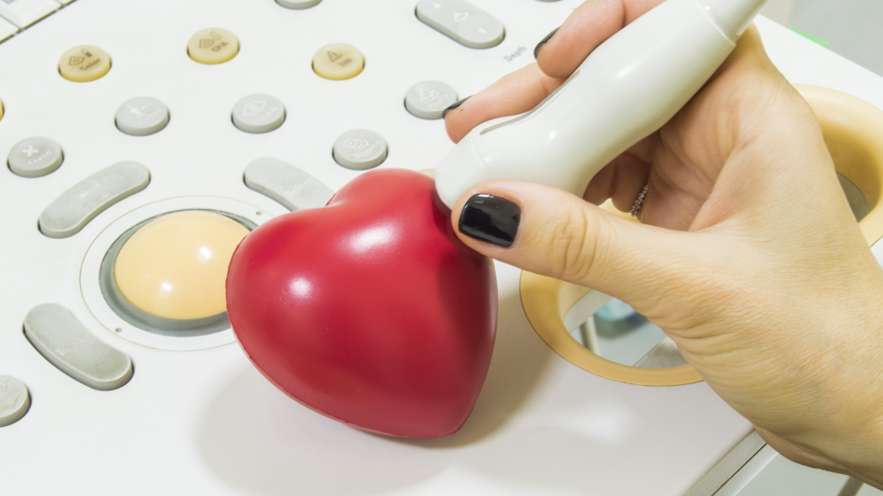
Echocardiography provides you with essential insights into your heart's function and detects potential pathologies by utilizing ultrasound waves to create images of cardiac structures. You can observe real-time blood flow dynamics, evaluate the heart chambers' size, and assess myocardial wall thickness. It also measures ejection fraction to gauge your heart's pumping efficiency and employs Doppler analysis to examine blood flow velocity, helping to identify abnormal patterns indicative of issues like valve dysfunctions or tissue damage. This non-invasive method offers a significant advantage for continuous monitoring and accurate diagnosis. Further exploration will enhance your understanding of your cardiac health.
Key Takeaways
- Echocardiography visualizes heart chambers and valves, assessing their structure and functionality.
- Doppler analysis measures blood flow velocity, detecting abnormal flow patterns indicative of pathologies.
- Wall thickness measurements identify conditions like hypertrophic cardiomyopathy and myocardial infarction.
- Chamber size assessment through 2D and 3D echocardiography provides essential volume measurements.
- Tissue damage evaluation quantifies infarcted tissue and assesses myocardial wall motion abnormalities.
Understanding Echocardiograms
Delving into echocardiograms, it's crucial you understand that this diagnostic tool utilizes ultrasound waves to create detailed images of your heart's structure and function. This technique enables healthcare providers to assess cardiac conditions accurately and non-invasively, making it a cornerstone in diagnostic cardiology.
To begin with, the echocardiogram's efficacy hinges on its ability to generate real-time images that reveal the dynamics of blood flow through the heart's chambers and valves. This is achieved through the Doppler effect, which detects the changes in frequency of the ultrasound waves as they bounce off moving objects, such as blood cells. This aspect is pivotal, as it provides insight into the velocity and direction of blood flow, helping diagnose conditions like valve disorders or congenital heart defects.
Furthermore, echocardiograms are instrumental in evaluating the heart's pumping capacity. By analyzing parameters such as ejection fraction, healthcare professionals can determine the percentage of blood that's pumped out of the ventricles with each contraction. This measurement is crucial for diagnosing and managing heart failure.
Moreover, the non-invasive nature of an echocardiogram makes it an invaluable tool for continuous monitoring of patient heart health, aiding in proactive management of various cardiac conditions. This capability ensures that you can provide compassionate and effective care to those in need.
Exploring Cardiac Anatomy
Building on the understanding of echocardiograms, let's now examine the detailed anatomy of the heart, a complex organ central to these diagnostic evaluations. You'll find that the heart isn't merely a muscular pump; it's an intricately structured entity composed of specialized tissues that coordinate the vital task of blood circulation.
The heart comprises four primary chambers: the right and left atria, and the right and left ventricles. The atria, located at the upper portion, serve as receiving areas for blood, while the ventricles, situated below, function as the main pumping compartments. The myocardium, the thick muscular layer of the heart wall, is critical for the contraction process, propelling blood through the circulatory system.
Moreover, the septum, a robust wall of muscle, divides the heart longitudinally, separating the left and right sides to prevent the mixing of oxygenated and deoxygenated blood. This division is essential for the efficient functioning of the heart and, consequently, the entire body.
As you delve deeper into cardiac anatomy via echocardiography, you'll also observe the coronary arteries and veins enveloping the heart surface. These vessels are paramount in delivering oxygen-rich blood to the myocardium, ensuring that it functions optimally under various physiological demands. Understanding this complex anatomy helps you appreciate the precision in diagnosing heart-related anomalies.
Assessing Valve Function
Transitioning from the broader aspects of cardiac anatomy, we now focus on assessing valve function, a critical component in maintaining unidirectional blood flow throughout the heart. Echocardiography offers you a dynamic tool for evaluating the structural integrity and functional performance of the heart valves. Each valve's ability to open and close properly is paramount in preventing regurgitation or stenosis, conditions that can lead to cardiac inefficiency and patient distress.
Using echocardiography, you're able to visualize the valves in real-time, assessing abnormalities in their morphology that could indicate pathologies like valvular insufficiency or prolapse. You'll examine the leaflets, assessing for thickening, calcification, or restricted movement, all of which can impair valve function. The annular dimensions are also crucial; dilatation here might suggest a failure in the supportive structure, prompting further investigation or intervention.
Moreover, the echocardiographic approach allows you to see the sequential timing of valve openings and closings within the cardiac cycle. This aspect is critical; even slight deviations from the norm can significantly impact cardiac output and overall circulatory efficiency. By mastering this evaluation, you enhance your ability to diagnose and manage potential cardiac issues, improving patient outcomes in your clinical practice.
Utilizing Doppler Analysis
In leveraging Doppler analysis, you'll measure the velocity of blood flow, a critical indicator of cardiovascular health.
This technique also allows for precise assessment of heart valve function, identifying abnormalities in flow patterns that might suggest valvular issues.
Blood Flow Velocity Measurement
Doppler analysis critically measures blood flow velocity, providing essential insights into cardiovascular function and anomalies. As you assess patients, understanding the rate and pattern of blood flow through their circulatory system is crucial.
This technique utilizes sound waves that reflect off moving blood cells. When these waves return to the echocardiography device, they've altered in frequency, dependent on the speed and direction of blood flow.
Heart Valve Function Assessment
Building on the understanding of blood flow velocity, let's examine how Doppler analysis aids in assessing the function of heart valves.
Doppler echocardiography provides critical insights by measuring the speed and direction of blood flow through cardiac valves. This technique detects abnormal flow patterns that may indicate stenosis or regurgitation, conditions where valves don't open fully or leak.
You'll analyze the spectral waveform obtained during Doppler assessment to identify these discrepancies. For instance, a high-velocity jet signals stenosis, whereas a backward flow suggests regurgitation.
Accurate readings are paramount for devising effective treatment plans. Therefore, mastering Doppler techniques will empower you to better serve patients by diagnosing and managing potential heart valve pathologies accurately and promptly.
Measuring Wall Thickness
As you assess myocardial infarction using echocardiography, measuring wall thickness is crucial for identifying areas of myocardial thinning and scarring. This parameter also helps in detecting hypertrophic cardiomyopathy, where increased wall thickness may indicate abnormal heart muscle growth.
Precise measurement techniques are essential to differentiate between pathological conditions and normal physiological variations.
Detecting Hypertrophic Cardiomyopathy
Echocardiography reliably measures ventricular wall thickness to detect hypertrophic cardiomyopathy, a critical indicator of this cardiac condition. By assessing the thickness of the heart's walls, you can identify abnormal myocardial hypertrophy, a hallmark of this disease. This technique involves high-resolution imaging that provides a detailed view of the myocardial structure, allowing you to discern even subtle deviations from normal thickness.
The precise measurement is crucial, as hypertrophic cardiomyopathy often leads to significant health risks, including heart failure and sudden cardiac death. Using echocardiography, you'll evaluate the septal and posterior wall dimensions, typically increased in this disorder. Recognizing these changes early helps tailor interventions that can mitigate complications, emphasizing your role in safeguarding cardiac health through vigilant monitoring and timely response.
Assessing Myocardial Infarction
Measuring wall thickness in myocardial infarction through echocardiography allows you to detect areas of diminished myocardial movement and thickening, indicative of possible tissue damage. This method aids in quantifying the extent of infarcted tissue, crucial for tailoring patient-specific therapeutic strategies.
By analyzing echocardiographic images, you can assess myocardial wall thickness variations and motion abnormalities across different cardiac cycles. This evaluation is critical as areas with increased wall thickness may suggest compensatory hypertrophy adjacent to infarcted zones. Conversely, thinning of the myocardial wall often signals severe myocardial damage.
Accurate interpretation of these findings enables you to provide targeted interventions, thereby improving patient outcomes and facilitating optimal recovery.
Evaluating Chamber Size
To accurately evaluate chamber size, cardiologists rely on specific echocardiographic techniques that quantify dimensions and volume. You'll find that two-dimensional (2D) echocardiography provides essential measurements of the left ventricle, right ventricle, and atria. By assessing these dimensions, you can determine if there are abnormalities like dilatation or hypertrophy, which are critical for diagnosing conditions such as cardiomyopathies or heart failure.
Moreover, 3D echocardiography offers even more detailed insights by enabling the calculation of chamber volumes and ejection fraction with higher accuracy and reproducibility than 2D methods. This technique involves acquiring a full volume dataset from which precise measurements can be derived, assisting in the management of various cardiac conditions and guiding therapeutic interventions.
It's crucial for you to understand the standard reference values for chamber sizes and how deviations from these norms can indicate pathological changes. Utilizing advanced quantification software, echocardiograms can be analyzed with high precision, providing you with reliable data to support your diagnostic decisions. This dedication to detail ensures that every patient you serve receives a diagnosis based on comprehensive and accurate cardiac assessment, ultimately enhancing patient care and outcomes.
Detecting Pericardial Effusion
When you assess for pericardial effusion using echocardiography, you're identifying any abnormal fluid accumulation around the heart.
This detection is crucial as it can significantly impact heart function by restricting cardiac output.
You'll rely on specific diagnostic imaging techniques to quantify the effusion and analyze its effects on heart dynamics.
Identifying Fluid Accumulation
Echocardiography frequently serves as a crucial tool in detecting pericardial effusion, enabling precise visualization of fluid accumulation around the heart. As you assess the echocardiographic images, you'll note the echogenic(bright) or anechoic(dark) space representing fluid in the pericardial cavity. This technique allows you to quantify the effusion, categorizing it as mild, moderate, or severe based on the volume of fluid observed.
You should also scrutinize the distribution of fluid, noting whether it's localized or circumferential. This detail aids in determining the potential need for intervention. By identifying these variations, you enhance your ability to tailor patient management strategies, ultimately aiming to alleviate suffering and improve cardiac outcomes for those in your care.
Impact on Heart Function
Pericardial effusion's impact on heart function can vary, often leading to compromised cardiac output as fluid pressure inhibits the heart's ability to pump efficiently.
You'll find the heart's chambers may become compressed, making it challenging for the ventricles to fill properly during diastole. This condition, termed cardiac tamponade, necessitates immediate intervention.
The increased intrapericardial pressure can progressively elevate, reducing venous return to the heart and consequently, decreasing stroke volume and cardiac output.
This pathophysiological cascade can lead to hypotension, tachycardia, and pulsus paradoxus, where there's a significant drop in blood pressure during inspiration.
Understanding these dynamics is crucial for timely and effective treatment, aiming to alleviate the effusion and restore cardiac function to support those in need.
Diagnostic Imaging Techniques
To accurately diagnose pericardial effusion, clinicians often rely on advanced imaging techniques, including echocardiography, which provides detailed visualizations of the heart's structure and function.
When you're using echocardiography, you're looking at a non-invasive method that utilizes ultrasound waves to create images of the heart. This method is crucial for identifying the accumulation of fluid in the pericardial cavity—a condition known as pericardial effusion.
The technique allows for the precise measurement of the effusion's volume and its hemodynamic impact. By assessing the size and distribution of the effusion, you can determine the urgency of intervention needed.
Furthermore, echocardiography helps in guiding pericardiocentesis, enhancing your ability to serve patients by providing targeted and effective treatments.
Identifying Artifacts
When interpreting echocardiographic images, you must distinguish real cardiac structures from artifacts to ensure accurate assessments. Artifacts in echocardiography can mimic pathologies, leading to diagnostic errors. You'll encounter several common types, including reverberation, side lobes, and mirror images.
Reverberation artifacts occur when ultrasound waves bounce between two strong reflectors, creating multiple, equally spaced echoes that can be mistaken for actual structures. To differentiate these, you should look for their characteristic linear, parallel appearance that doesn't conform to anatomical expectations.
Side lobe artifacts arise from the transmission of secondary ultrasound beams that radiate out from the main beam. These can cause structures outside the primary ultrasound field to appear within it, potentially leading to misinterpretation of tissue density or presence of masses. Adjusting the transducer's angle or frequency often helps minimize side lobes, enhancing your image clarity.
Mirror image artifacts are produced when ultrasound waves reflect off a strong reflector, like the diaphragm, causing structures to appear duplicated on the opposite side. Recognizing the anatomical implausibility of these mirrored structures is key to identifying this artifact.
Careful analysis of these artifacts is crucial. Your ability to accurately identify them directly impacts the reliability of your diagnostic interpretation, ultimately affecting patient care.
Generating Diagnostic Reports
You must ensure that echocardiographic reports are meticulously crafted to accurately reflect findings and guide clinical decision-making. Each report should succinctly detail the patient's cardiac structure and function, clearly delineating measurements like ventricular dimensions, wall thickness, and ejection fraction. It's crucial to annotate any pathological deviations, such as hypertrophy or dilatation, with precision.
When describing abnormalities, you'll need to integrate specific echocardiographic findings with potential pathophysiological implications. For instance, the presence of a septal defect or valvular insufficiency must be quantified and described in terms of severity and probable impact on cardiac function. Utilize standardized terminology and classifications to enhance the clarity and utility of your reports. This ensures consistency across different practitioners and aids in the formulation of a cohesive treatment strategy.
Moreover, it's vital to include a detailed description of the methods used, such as the type of echocardiogram ( transthoracic or transesophageal) and any specific imaging techniques employed. This information is essential for verifying the reliability of the findings and facilitating subsequent comparative assessments.
Correlating Clinical Data
Building on the meticulous documentation of echocardiographic findings, it's imperative to correlate these details with broader clinical data to enhance diagnostic accuracy and patient management. You'll need to integrate echocardiographic results with patient history, presenting symptoms, and other diagnostic tests. This comprehensive approach ensures a more accurate assessment of cardiac function and potential pathologies.
For instance, if echocardiography shows left ventricular hypertrophy, you should examine hypertension or aortic valve disease as potential underlying causes. Cross-referencing the echocardiographic data with patient-reported symptoms and blood pressure readings is crucial. It helps you differentiate between possible etiologies and guide the therapeutic approach.
Moreover, in cases of suspected cardiac dysfunctions like heart failure, correlating echocardiographic findings such as ejection fraction and ventricular dimensions with clinical indicators like edema, dyspnea, and fatigue is essential. This correlation aids in staging the disease and tailoring patient-specific management strategies.
Conclusion
In summary, echocardiography is your window into the heart's detailed mechanics. By assessing valve functionality, measuring wall thickness, and utilizing Doppler analysis, you're equipped to detect subtle abnormalities before they escalate.
Remember, 'a stitch in time saves nine'; early detection through echocardiography can prevent severe cardiac complications. Correlating these findings with clinical data ensures a comprehensive diagnostic report, enhancing patient management strategies.
Always scrutinize for artifacts to maintain diagnostic accuracy.


