Blog and News

Echocardiography lets you peer into the intricate workings of your heart, revealing crucial details that influence the management of heart failure. By measuring your ejection fraction, this technique quantifies how much blood your left ventricle pumps out with each beat, helping to pinpoint stages of heart failure. It goes further by assessing ventricular function, detecting any dilation, and evaluating wall motion abnormalities. This data is vital for adjusting your treatment plan effectively. Through non-invasive imaging, echocardiography offers a comprehensive view that guides clinical decisions and improves patient outcomes. Exploring these diagnostic insights further can be crucial in managing your heart health more proactively.
Listen to the Article
Key Takeaways
- Echocardiography measures ejection fraction, revealing how much blood the heart pumps, aiding in diagnosing heart failure.
- It identifies wall motion abnormalities, pinpointing areas of the heart muscle that are underperforming or damaged.
- Regular echocardiographic assessments monitor changes in heart function and effectiveness of treatments over time.
- The technique detects ventricular dilation and volume overload, indicators of worsening heart failure.
- Doppler ultrasound within echocardiography assesses blood flow velocities, helping calculate cardiac output for clinical decision-making.
Understanding Echocardiography Basics
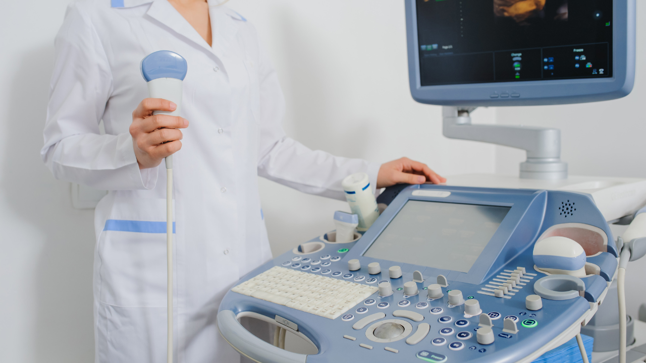
Echocardiography, often used to assess heart function, employs sound waves to create detailed images of your heart's structure and motion. This imaging tool is crucial for diagnosing various cardiac conditions, guiding treatment decisions, and monitoring therapy effectiveness. When you perform echocardiography, you're not just operating a machine; you're providing a vital service that can profoundly impact patient care.
As a healthcare professional, you'll utilize two main types of echocardiography: transthoracic (TTE) and transesophageal (TEE). TTE is non-invasive and widely used; it involves placing the transducer on the patient's chest where it emits sound waves that bounce off the heart, producing images for analysis. TEE, however, involves inserting a probe into the esophagus to get closer to the heart, providing clearer, more detailed pictures. This method is particularly useful when TTE doesn't yield sufficient information or when you're assessing certain conditions such as atrial fibrillation or congenital heart defects.
Understanding these techniques allows you to tailor your approach to each patient's needs, optimizing diagnostic accuracy while ensuring their safety and comfort. Each scan you conduct offers critical insights that aid in the accurate assessment of heart health, enhancing your ability to serve and support recovery effectively.
Measuring Ejection Fraction
While assessing heart function, measuring ejection fraction is crucial as it quantifies how much blood the left ventricle pumps out with each contraction. This metric is vital for you, as a healthcare provider, to determine the severity of heart failure in your patients and to guide therapeutic decisions effectively.
Ejection fraction (EF) is typically expressed as a percentage, indicating the proportion of blood pumped out of the ventricle with each heartbeat in relation to the total volume of blood in the ventricle at the end of filling. A normal EF ranges from 55% to 70%, but lower values suggest impaired cardiac function, which might require intervention.
To keep you engaged and informed, here are key points about EF measurement using echocardiography:
- Accuracy and Non-Invasiveness: Echocardiograms provide a precise EF measurement without needing invasive procedures.
- Diagnostic Benchmark: EF is a critical indicator in diagnosing heart failure and other cardiac conditions.
- Progress Monitoring: Regular EF assessments can help monitor the effectiveness of heart failure treatment.
- Risk Stratification: EF aids in assessing patient risk and tailoring individual management plans.
Mastering EF measurement enhances your ability to serve patients with heart-related ailments effectively, ensuring they receive the most informed care possible.
Assessing Heart Muscle Function
Assessing heart muscle function is critical in understanding the full scope of heart failure.
You'll need to evaluate ventricular contraction efficiency to gauge how well the heart pumps with each beat.
Additionally, identifying regional wall motion abnormalities can help pinpoint areas of the heart that aren't functioning properly, while accurately measuring ejection fraction provides a quantitative assessment of cardiac output.
Evaluating Ventricular Contraction Efficiency
To gauge the efficiency of ventricular contraction, clinicians often rely on specific echocardiographic parameters that measure heart muscle function directly. Understanding these metrics is crucial for diagnosing and managing heart failure effectively.
Here are key parameters to consider:
- Ejection Fraction (EF): Measures the percentage of blood ejected from the ventricle during each contraction.
- Fractional Shortening: Assesses the reduction in diameter of the ventricle during systole compared to diastole.
- Stroke Volume: Quantifies the volume of blood pumped out of the ventricle with each heartbeat.
- Cardiac Output: Calculates the amount of blood the heart pumps through the circulatory system in one minute.
Identifying Regional Wall Motion
Building on our understanding of ventricular contraction efficiency, we now explore how echocardiography can help detect abnormalities in regional wall motion to assess heart muscle function.
This tool provides a dynamic visual representation of how different sections of the heart wall move during each heartbeat. When you observe an area that isn't contracting or relaxing properly, it could indicate localized damage, such as from a previous heart attack or ongoing ischemic disease.
You'll assess the thickness and motion of the myocardial segments during systole and diastole, identifying hypokinetic (reduced movement), akinetic (no movement), or dyskinetic (abnormal movement) areas. This precise evaluation is crucial for diagnosing the extent and specific location of heart muscle dysfunction, enabling targeted treatments to restore cardiac health.
Measuring Ejection Fraction Accurately
Echocardiography provides a reliable method for measuring your heart's ejection fraction, a critical indicator of its pumping efficiency. This measurement is vital for diagnosing and managing heart failure, ensuring you can offer the best care to those in need.
- High Resolution Imaging: Captures detailed views of the heart chambers to assess real-time cardiac function.
- Temporal Accuracy: Offers precise timing of cardiac cycles to evaluate systolic and diastolic performance.
- Reproducibility: Ensures consistent measurements across different exams and operators, crucial for monitoring disease progression.
- Non-Invasive Procedure: Safeguards patient comfort and reduces risk, facilitating easier, repeated assessments.
Analyzing Cardiac Output
Analyzing cardiac output effectively helps you understand the heart's efficiency in pumping blood throughout the body. As a healthcare provider, your ability to assess this key parameter using echocardiography is crucial in managing patients with heart failure. Cardiac output, quantified as the volume of blood the heart ejects in one minute, is a fundamental measure of cardiac function.
To accurately evaluate cardiac output, you'll rely on echocardiographic techniques such as Doppler ultrasound to measure blood flow velocities across the cardiac valves. You must integrate these velocity data with the heart valve areas during various phases of the cardiac cycle. This integration allows you to calculate stroke volume —the amount of blood pumped with each heartbeat. Multiplying the stroke volume by the heart rate gives you the cardiac output.
Understanding these values in real-time can guide your clinical decisions, especially in critical care settings. For instance, a decreased cardiac output may indicate worsening heart failure, prompting immediate therapeutic interventions. Conversely, monitoring improvements in cardiac output can affirm the effectiveness of your treatment regimen, providing both you and your patient with valuable feedback.
Mastering these assessment techniques not only enhances your diagnostic capabilities but also profoundly impacts patient care, emphasizing your role in their health journey.
Detecting Ventricular Dilation
Detecting ventricular dilation is crucial for evaluating the progression of heart failure in patients. As a healthcare provider, you're well aware that the heart's ability to pump effectively is paramount, and any changes in the size of the ventricles can be a telltale sign of worsening or improving condition. Echocardiography stands as a non-invasive, reliable method to assess these changes, providing you with the necessary information to adjust treatment plans effectively.
Consider these key aspects when using echocardiography to detect ventricular dilation:
- Measurement of Chamber Size: Accurate quantification of the dimensions of the ventricles is essential, focusing on end-diastolic and end-systolic volumes.
- Assessment of Ventricular Function: Evaluate the ejection fraction to understand how much blood the ventricle pumps out with each contraction, which can be indicative of dilation.
- Detection of Volume Overload: Identifying signs of increased preload that can stretch the ventricle, leading to dilation.
- Longitudinal Monitoring: Regular scanning to track changes over time, providing insight into the progression or improvement of the patient's heart function.
Identifying Wall Motion Abnormalities
Beyond measuring ventricular size, you must also assess wall motion abnormalities to fully evaluate heart function. This critical aspect involves examining the movement of the myocardial walls during cardiac cycles. In echocardiography, you'll use techniques like M-mode and 2D echocardiography to detect these abnormalities, which are often indicative of ischemic heart disease or cardiomyopathies.
You'll look for hypokinetic, akinetic, or dyskinetic areas within the myocardial tissue. Hypokinetic sections exhibit reduced movement, often signaling reduced blood flow or prior myocardial infarction. Akinetic areas, showing no movement, and dyskinetic regions, which move paradoxically during systole, are severe and require immediate attention. These findings can guide you towards specific therapeutic interventions and influence the prognosis.
Moreover, it's crucial to quantify the degree of these abnormalities accurately. Echocardiography allows for detailed measurement of wall thickening and excursion, which are vital metrics in determining the extent of damage or dysfunction. Your ability to interpret these signs not only aids in diagnosis but significantly impacts the management and monitoring of patients with heart failure.
Exploring Diastolic Dysfunction
As you explore diastolic dysfunction through echocardiography, you'll first focus on identifying signs of impaired ventricular relaxation, a key indicator of this condition.
You'll then assess ventricular relaxation techniques to gauge their efficiency and detect any abnormalities in the heart's ability to fill during diastole.
Identifying Diastolic Dysfunction
To explore diastolic dysfunction, echocardiography evaluates the heart's ability to relax and fill during the diastolic phase. This crucial assessment helps you pinpoint the subtle deviations that may indicate early or evolving heart issues in patients who rely on your expertise for their health and well-being.
- E/A Ratio Analysis: This involves measuring the ratio of early (E) to late (A) ventricular filling velocities, a key indicator of diastolic function.
- Pulmonary Vein Flow: Examining the flow patterns in pulmonary veins can provide insights into atrial pressures and diastolic performance.
- Left Atrial Size Measurement: An enlarged left atrium often suggests chronic diastolic dysfunction.
- Tissue Doppler Imaging: This technique assesses myocardial velocities, offering a detailed view of the regional diastolic function.
Assessing Ventricular Relaxation
Assessing ventricular relaxation is crucial for identifying the severity and progression of diastolic dysfunction in your patients.
Utilizing echocardiography, you can measure the myocardial relaxation and filling velocities which are key indicators of ventricular compliance.
Specifically, the early diastolic velocity (E') obtained via tissue Doppler imaging provides a direct quantification of myocardial relaxation.
A reduced E' signifies impaired relaxation, a hallmark of early diastolic dysfunction.
Analyzing Filling Pressures
Understanding filling pressures through echocardiography is pivotal for diagnosing the extent of diastolic dysfunction in your patients. By assessing how the heart fills with blood during diastole, you can determine the pressure within the heart chambers without invasive procedures. This insight is crucial for tailoring the most effective treatment plans.
- E/A Ratio Analysis: Evaluates early (E) and late (A) ventricular filling velocities.
- Deceleration Time: Measures the rate of decline in early filling, providing insights into ventricular relaxation.
- Pulmonary Venous Flow: Assesses the systolic and diastolic flow velocities in pulmonary veins, indicating left atrial pressures.
- Left Atrial Volume Index: Helps estimate left atrial pressure, especially in chronic diastolic dysfunction.
This approach ensures a comprehensive understanding, allowing you to optimize care for those you serve.
Impact on Prognosis and Management
Echocardiography significantly influences the prognosis and management strategies for patients with heart failure. By providing detailed visualization of cardiac structure and function, you can more accurately assess the severity and progression of the disease. This tool enables you to detect subtle changes in ventricular function, valvular abnormalities, and other cardiac complications that would otherwise go unnoticed with conventional diagnostic methods.
Through echocardiographic evaluation, you're also able to monitor the effectiveness of therapeutic interventions. Adjustments in treatment plans can be made based on real-time feedback regarding how a patient's heart is responding to current therapies. This is crucial in preventing the progression of heart failure and improving patient outcomes.
Moreover, echocardiography assists in risk stratification. By identifying patients at higher risk of adverse events, you can tailor more aggressive treatment strategies to those who need it most, while avoiding unnecessary interventions in lower-risk individuals. This judicious use of resources not only optimizes patient care but also reduces the burden on healthcare systems.
In your role, utilizing echocardiography to its full potential ensures that you're not just treating heart failure; you're managing it with precision, enhancing both the quality and the length of life for your patients.
Conclusion
As you delve deeper into the shadows of heart failure with echocardiography, each scan becomes a critical reveal. Picture the heart's chambers expanding, the ejection fraction faltering—subtle cues to impending danger.
Visualize muscles struggling, wall motions betraying distress, each image sharpening your clinical acumen. Embrace this tool, for it not only maps the present but also foretells the heart's resilience or its surrender.
Your expertise, armed with these insights, becomes pivotal in steering patient outcomes from uncertainty to calculated precision.
Services
Contact Details
Address: 1971 Gowdey Road,
Naperville, IL 60563
Phone: 630-416-1300
Fax:
630-416-1511
Email: info@foxvalleyimaging.com
© Copyright 2023 Fox Valley Imaging, Inc..


