Blog and News
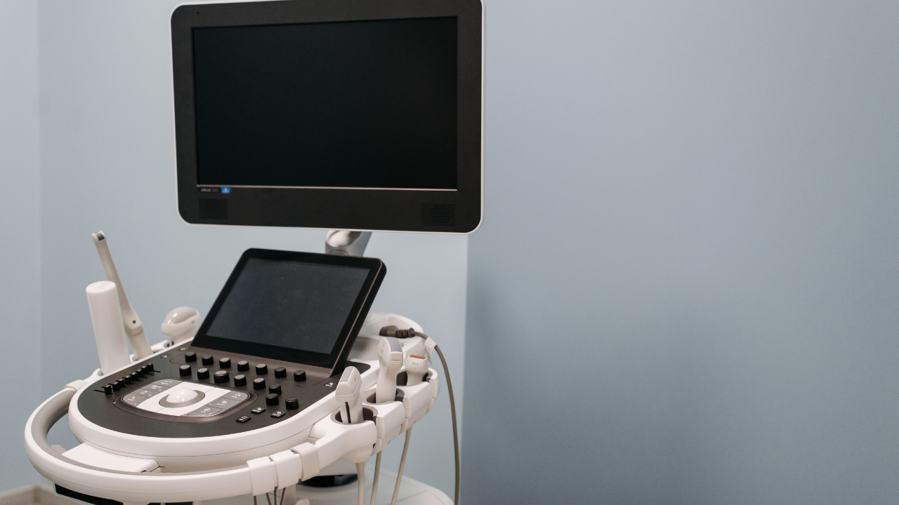
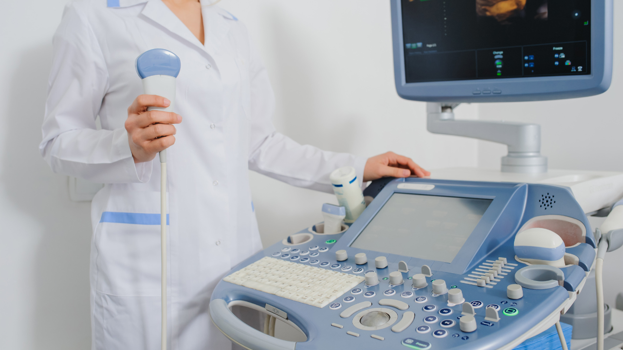
You can rely on ultrasound for early cancer detection due to its high-resolution imaging and ability to visualize abnormal tissue structures precisely. It accurately measures tumor dimensions, assesses vascularity, and characterizes echotextures to differentiate benign from malignant masses. Doppler imaging proves invaluable in evaluating blood flow and detecting subtle tissue changes. Ultrasound-guided biopsies utilize real-time imaging to enhance needle placement precision, reducing sampling errors. Furthermore, it effectively evaluates lymph nodes, distinguishing between reactive and metastatic nodes with high sensitivity and specificity. For a comprehensive exploration of its capabilities in different cancer types, there's much more to discover.
Listen to the Article
Key Takeaways
- Ultrasound provides high-resolution imaging, enabling the precise visualization of abnormal tissue structures for early cancer detection.
- Doppler imaging enhances the assessment of blood flow, aiding in the differentiation between benign and malignant masses.
- Real-time ultrasound imaging guides biopsy needles accurately, reducing sampling errors and improving diagnostic yield.
- Ultrasound effectively evaluates lymph node characteristics, distinguishing between reactive and metastatic nodes with high sensitivity and specificity.
- Tissue density analysis through ultrasound helps identify hypoechoic regions, which are indicative of malignancy, aiding in accurate diagnoses.
Role of Ultrasound in Cancer Detection
Ultrasound technology, with its high-resolution imaging capabilities, plays a pivotal role in the early detection of various cancers by allowing for the precise visualization of abnormal tissue structures.
Evaluating Tumors With Ultrasound
When evaluating tumors with high-frequency ultrasound, clinicians can:
- Precisely measure tumor dimensions
- Assess vascularity
- Characterize the echotexture to differentiate between benign and malignant masses
Doppler imaging enhances the assessment of blood flow within the tumor, providing critical insights into neovascularity.
High-resolution imaging allows you to detect subtle changes in tissue architecture, ensuring accurate and timely diagnoses, thus improving patient outcomes and advancing clinical care.
Ultrasound for Biopsy Guidance
Clinicians leverage real-time ultrasound imaging to guide biopsy needles with precision, enhancing the accuracy of tissue sampling and minimizing complications.
You utilize high-frequency sound waves to visualize the target lesion, ensuring optimal needle placement.
This method significantly reduces the risk of sampling error and procedural morbidity, thereby improving diagnostic yield.
Ultrasound-guided biopsies are indispensable in oncologic care, ensuring patients receive timely and accurate diagnoses.
Analyzing Tissue Density
High-resolution imaging not only aids in guiding biopsies but also plays a pivotal role in assessing tissue density to differentiate between benign and malignant lesions.
You'll leverage ultrasound's ability to measure acoustic impedance variations, identifying hypoechoic regions indicative of malignancy.
Lymph Node Evaluation
Leveraging ultrasound technology, you can effectively evaluate lymph node characteristics, such as size, shape, and internal architecture, to distinguish between reactive and metastatic nodes.
Studies show high sensitivity and specificity in detecting malignancy, aiding in early intervention. Utilizing Doppler imaging enhances vascularity assessment, providing critical diagnostic information.
This method ensures precise, non-invasive evaluation, aligning with your commitment to early cancer detection and patient care.
Detecting Liver Cancer Early
Early detection of liver cancer is crucial and relies on ultrasound technology to pinpoint subtle abnormalities in hepatic tissue. This method significantly enhances patient prognosis by excelling in identifying small lesions and vascular anomalies through the use of high-frequency sound waves.
Empirical studies have demonstrated sensitivity rates surpassing 80% in high-risk populations, enabling timely intervention. This non-invasive approach aligns with your goal of providing superior patient care efficiently.
Ultrasound in Breast and Prostate Cancer
When considering ultrasound for breast and prostate cancer, you'll find it excels in screening accuracy rates, particularly in dense breast tissue and early-stage prostate tumors.
Advanced tumor visualization techniques, like Doppler ultrasound and elastography, enhance the detection of malignancies.
Compared to other modalities, ultrasound offers superior diagnostic efficacy in specific patient populations, reducing the need for invasive biopsies.
Screening Accuracy Rates
Evaluating the screening accuracy rates of ultrasound for detecting breast and prostate cancer reveals significant variations influenced by factors such as tumor size, location, and operator proficiency. Studies indicate that sensitivity ranges from 60% to 80% for breast cancer, while prostate cancer detection varies widely, often dependent on transrectal versus transabdominal approaches.
Consistent training and experience markedly improve diagnostic accuracy, ultimately enhancing patient outcomes.
Tumor Visualization Techniques
Understanding the nuances of tumor visualization techniques is vital, as ultrasound's ability to differentiate tissue characteristics significantly impacts the early detection of breast and prostate cancers.
By employing high-frequency sound waves, you can identify hypoechoic regions indicative of malignancy. Doppler ultrasound further enhances detection by revealing abnormal vascular patterns.
This precision aids in early intervention, significantly improving patient outcomes and advancing your commitment to serving others.
Comparative Diagnostic Efficacy
Ultrasound's diagnostic efficacy in breast and prostate cancer hinges on its ability to provide real-time, high-resolution imaging that distinguishes malignant from benign tissue with remarkable accuracy.
In breast cancer, ultrasound effectively identifies lesions, particularly in dense breast tissue, while in prostate cancer, it aids in detecting abnormal growths and guiding biopsies.
Both applications enhance early detection, offering significant clinical value in patient outcomes.
Frequently Asked Questions
What Are the Limitations of Ultrasound in Cancer Detection?
Ultrasound's limitations include operator dependency, difficulty in imaging dense tissues, and limited resolution for small lesions. You might miss early-stage tumors, necessitating additional diagnostic tools for comprehensive cancer detection and accurate diagnosis.
How Does Ultrasound Compare to MRI in Detecting Cancer?
When comparing ultrasound to MRI in cancer detection, you'll find MRI offers superior soft tissue contrast and specificity. However, ultrasound is more accessible, cost-effective, and valuable for guiding biopsies, particularly in resource-limited settings.
Can Ultrasound Detect All Types of Cancer?
You might think ultrasound detects every cancer type, but it's not a catch-all solution. While effective for certain cancers like breast and thyroid, its sensitivity varies. Comprehensive diagnosis often requires combining modalities for accurate results.
Are There Risks Associated With Frequent Ultrasound Screenings?
Frequent ultrasound screenings carry minimal risks, primarily thermal and mechanical effects. However, studies show these risks are negligible compared to benefits, especially when early cancer detection can significantly improve patient outcomes. Always prioritize evidence-based guidelines.
How Should Patients Prepare for an Ultrasound Screening?
You should fast for 8-12 hours and wear loose clothing for abdominal ultrasounds. Hydrate well if a pelvic ultrasound is required. Follow these protocols to ensure optimal imaging and accurate diagnostic results.
Conclusion
In a nutshell, ultrasound's ability to detect and diagnose cancer early is like a vigilant sentinel, providing crucial insights.
By evaluating tumors, guiding biopsies, and analyzing tissue density, it delivers precise, real-time data. Its prowess in assessing lymph nodes, liver, breast, and prostate cancers is backed by robust evidence, showcasing its indispensable role in oncology.
Trust in ultrasound, and you're harnessing a powerful ally in the battle against cancer.


