Blog and News

How Ultrasound Can Also Be Used To Diagnosis Health Problems
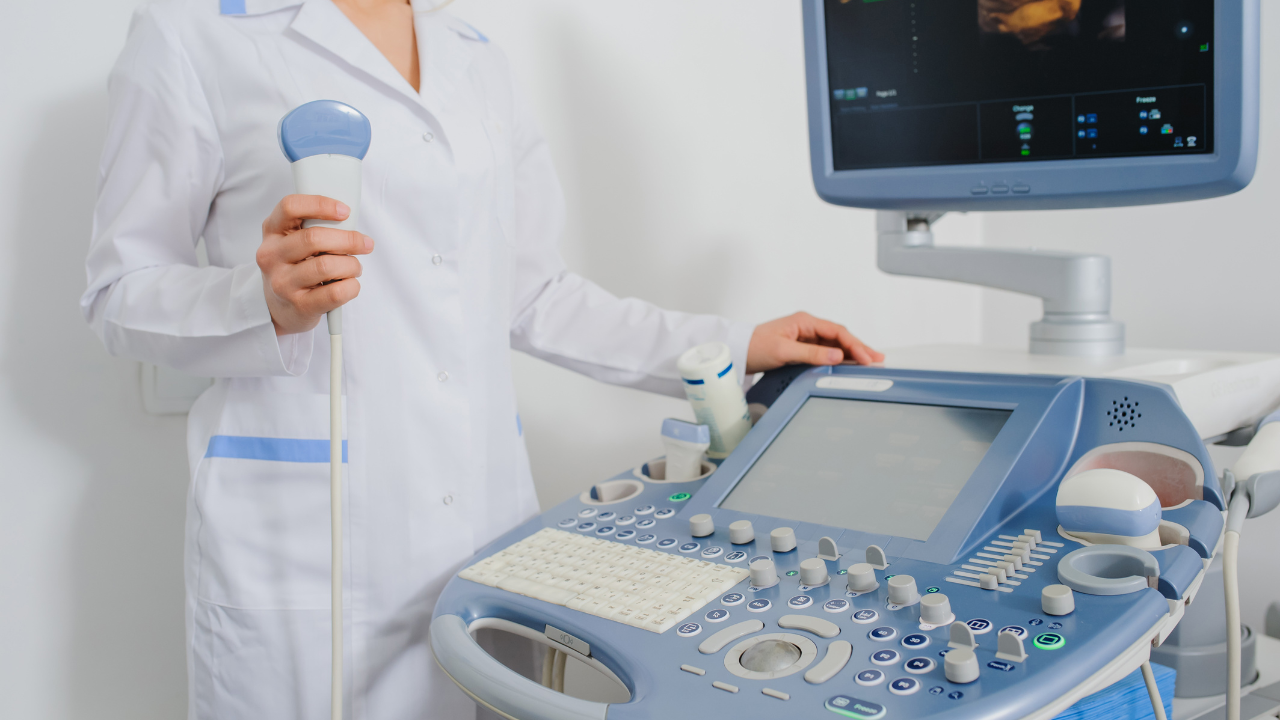
Ultrasound imaging, also called ultrasound scanning or sonography, involves exposing part of the body to high-frequency sound waves to produce pictures of the inside of the body. Ultrasound exams do not use ionizing radiation like an x-ray does. Because ultrasound images are captured in real-time, they can show the structure and movement of the body’s internal organs, as well as blood flowing through blood vessels.
Ultrasound imaging is usually a painless medical test that helps physicians diagnose and treat medical conditions. Conventional ultrasound displays the images in thin, flat sections of the body.
Ultrasound examinations can help to diagnose a variety of conditions and to assess organ damage following illness. Ultrasound is used to help physicians diagnose symptoms such as:
- pain
- swelling
- infection
Ultrasound imaging is based on the same principles involved in the sonar used by bats, ships and fishermen. When a sound wave strikes an object, it bounces backward, or echoes. By measuring these echo waves it is possible to determine how far away the object is and its size, shape, consistency (whether the object is solid, filled with fluid, or both) and uniformity.
In medicine, ultrasound is used to detect changes in appearance and function of organs, tissues, or abnormal masses, such as tumors.
In an ultrasound examination, a transducer sends the sound waves and also records the echoing waves. When the transducer is pressed against the skin, it directs a stream of inaudible, high-frequency sound waves into the body. As the sound waves bounce off of internal organs, fluids and tissues, the sensitive microphone in the transducer records tiny changes in the sound’s pitch and direction. These signature waves are instantly measured and displayed by a computer, which in turn creates a real-time picture on the monitor.
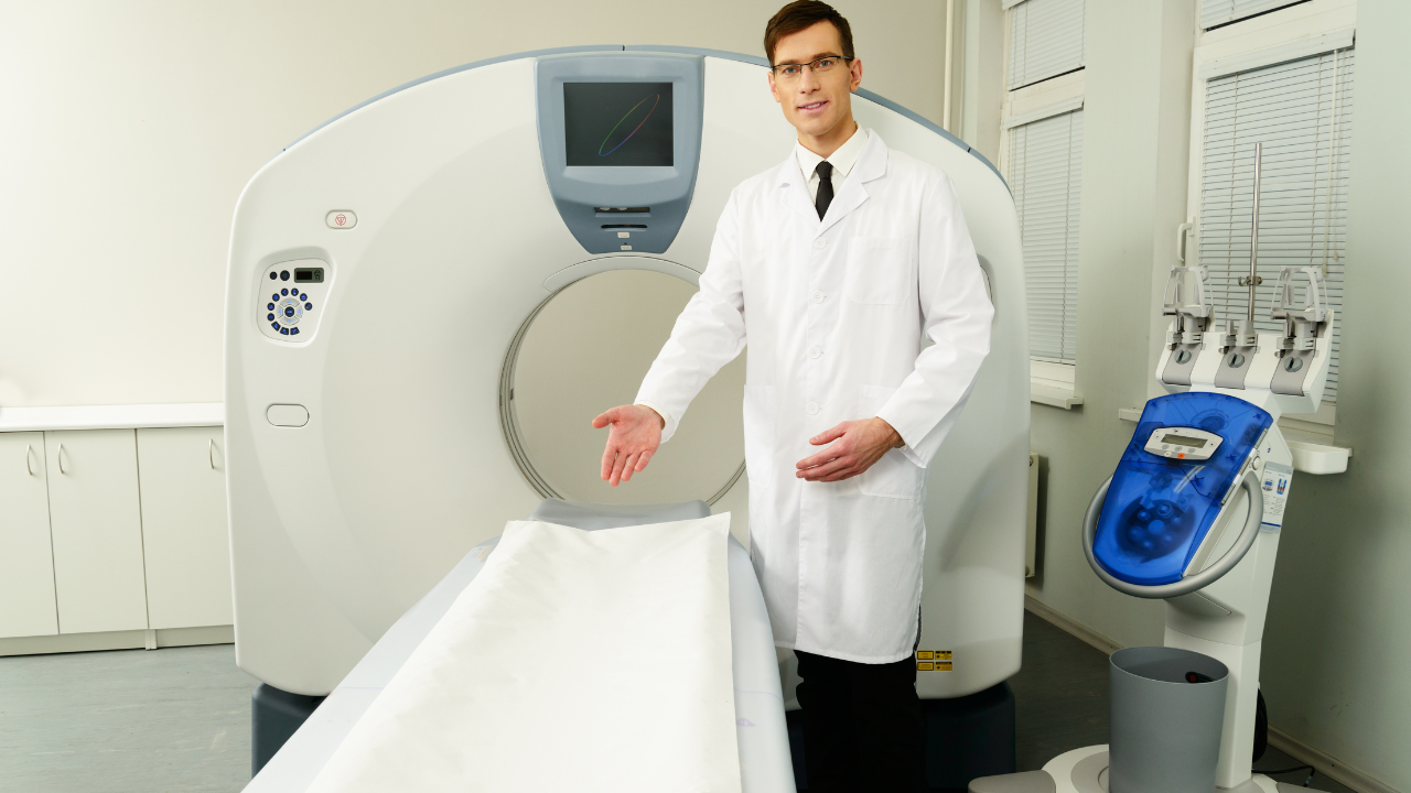
A radiologist, a physician specifically trained to supervise and interpret radiology examinations, will analyze the images and send a signed report to your primary care or referring physician, who will share the results with you. In some cases the radiologist may discuss preliminary results with you at the conclusion of your examination.