Blog and News

MRI: A Guided Tour
MRI scanners, like X-rays and CT scanners, are basically machines doctors use to take pictures of your insides so that they can figure out what’s ailing you. But MRI doesn’t involve ionizing radiation, as do X-rays and CT scans.
Rather, MRI takes advantage of something you have plenty of in your body: water. It is far more flexible than X-rays and CT scans can generate three-dimensional images in any orientation and at any depth in the body. The upshot is that MRI, for most applications, is far superior to other imaging tools in providing non-invasive images (and even chemical information) at high resolution.
While X-rays remain useful for looking at bones, MRI scans are the diagnostic tool of choice for soft tissue — organs, ligaments, the circulatory system and (as you know) the spinal column and cord. They help physicians identify multiple sclerosis, tumors, tendonitis, strokes, and many other conditions.
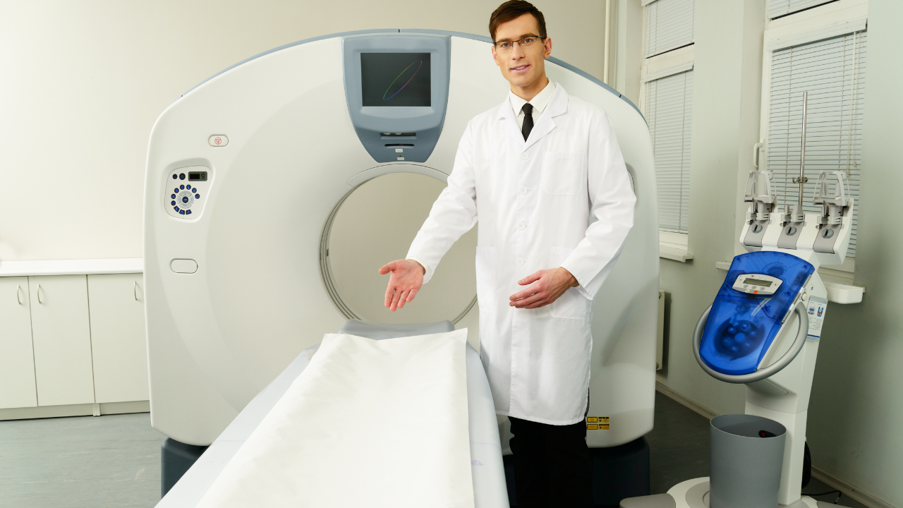
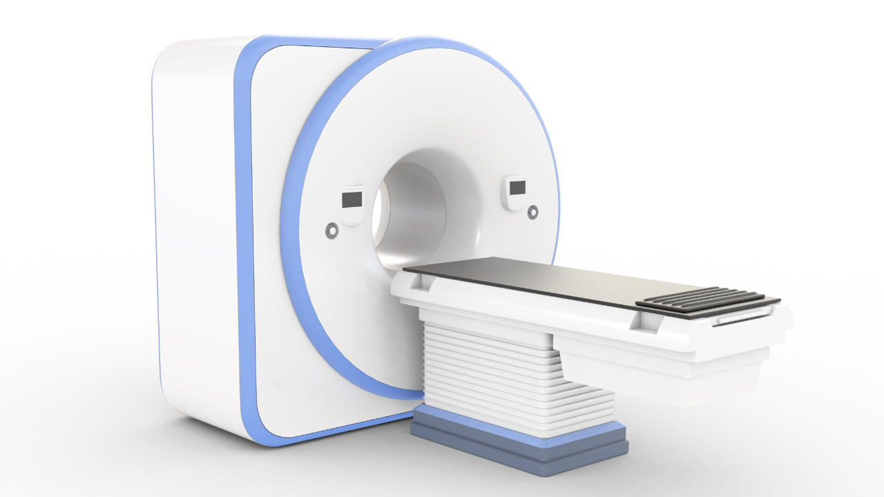
Let’s start with a little tour of the metallic cylinder surrounding you (it’s called a bore, technically). You’re in the center of a tremendous magnet, weighing tens of thousands of pounds and differing from the little magnets on your fridge in two fundamental ways. First, those fridge decorations are permanent magnets made of alloys. The MRI magnet surrounding you, on the other hand, is a superconducting magnet; it conducts electricity, thereby creating a magnetic field. Secondly, your fridge magnet has a fraction of the power of the one you’re in.
By now the technologist in the control room, who is talking you through the exam via an intercom, has started to place you into the main magnetic field in your scanner. The field is running horizontally through the bore from your head to your toes (or vice versa, depending on your position). Because your spine is what she’s interested in, you’re being positioned so that part of your back is in the middle (or isocenter) of the field.
Now that the magnet has gotten the hydrogen protons lined up at attention, the scanner is ready to subject them to the next step, the one that will result in an actual signal.
You’ve probably been wondering about that coil the technologist placed under your back. Essentially, it’s a radio transceiver, also called an RF coil, which can communicate with your hydrogen atoms via radio frequency (RF) waves.
Your technologist is using that coil to send RF pulses at your spine. They are precisely timed (taking into account the type of tissue targeted and the fact that just the hydrogen atoms are of interest) to achieve the effect we’re about to describe.
The signal gets turned into an electric current, which the scanner digitizes. Different types of MRIs display this data differently, but in any case you get a variety of shades of grey that reflect the different densities. In some scans, the weaker the signal, the darker that part of the image will be. So bone will be fairly dark, while fat will be light.
Believe it or not, MRI scans can display more than 250 distinct shades of grey, each reflecting slight variations in tissue density or water content. It is in those subtle shades that radiologists unlock the secrets of the tissues. For example, abnormal tissue, such as a brain tumor, will look different than the normal tissue surrounding it. The technologist and radiologist have the ability to alter imaging parameters (like the timings of the RF pulse and gradients) to emphasize areas of injury or disease or to acquire higher image resolutions.