Blog and News
Ultrasound Image Quality Improvements
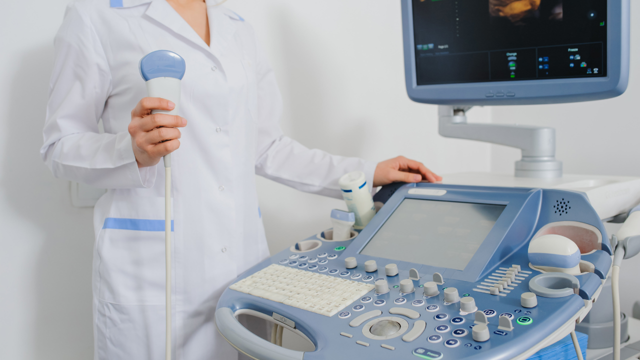
Today, ultrasound technology is producing images with much higher resolution than in the past. This allows doctors to see with much sharper definition. “Everyone is used to ultrasound pictures being fuzzy,” said Tomo Hasegawa, director, ultrasound business unit, Toshiba America Medical Systems. With enhancements in computer technology doing real-time processing, we’re starting to get images that are so clear, people don’t even realize it’s ultrasound”.
The principal reason why there has been such a great improvement in ultrasound equipment is because of the many recent upgrades to the technology. For instance, there has been a tremendous improvement in transducer sensitivity and beamformer. Also, the speed of processing and the final data display quality have been greatly improved. Because of these improvements, physicians are now able to distinguish details that are much smaller and a great deal deeper than was possible before these improvements. For example, you can now see the flow in vessels that are as tiny as 2 mm in diameters, such as what is found in structures like the lymph nodes and the kidneys.
Following are the current trends in medical imaging:
- Technologies that will provide better management of the dosage and exposure to ionizing radiation
- Solutions will be transportable, mobile and wireless
- Capabilities to evaluate tissue stiffness using elastography
- Technologies resulting in improved patient comfort
- Imaging equipment that is easy to use will become commonplace
Ultrasound is able to accomplish every one of these requirements.
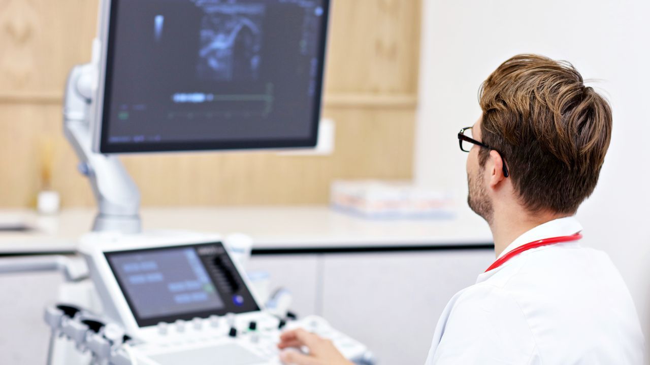
Whether it is for general imaging, ob/gyn or cardiac use, ultrasound has evolved to handle all types of examinations. Over time, as ultrasound has become more complex, users have become specialists who are more and more proficient in managing the system. At the same time, the miniaturization trend in ultrasound equipment is rapidly facilitating the ability to extend point-of-care usage, such as when the clinician is able to come to a diagnosis at the patient’s bedside using ultrasound for immediate diagnosis.
Many new ways to utilize ultrasound are quickly becoming available as the market expands into new areas. Ultrasound is now being used in breast imaging and biopsies, cardiology, head and neck surgery, gastroenterology, pulmonology, anesthesiology, ophthalmology, neurology, neonatology, vascular imaging, urology, emergency medicine and musculoskeletal exams and other possibilities.
At Fox Valley Imaging, we will always integrate the latest imaging advances in technology. Our 3T systems offer higher resolutions which help prevent misdiagnosis due to substandard older technologies found in many locations. We are leading the way to a more accurate diagnosis.