Blog and News

Understanding the Difference Between a Single Slice CT and a Multi-Slice CT
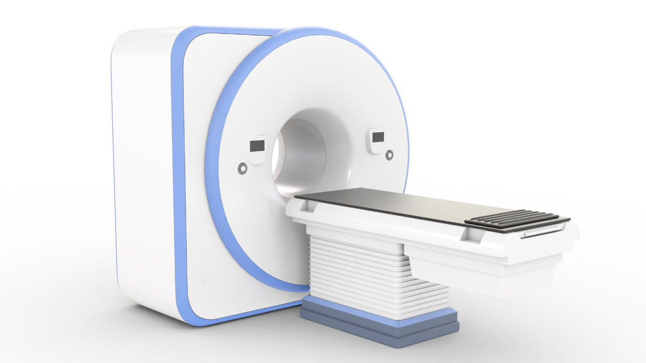
Multi-Slice CT Scanners
Different Types of CT Scans
In order to understand the different types of CT imaging scans, you must first understand the meaning of the word “tomography.” This Greek word comes from two distinct words “tomos” and “graphe.” “Tomos” means “section or slice” while “graphe” means “drawing.”
When a patient passes through the CT, the circular opening rotates and takes a series of x-rays. Each rotation takes approximately 1 second. During the rotation, beams of radiation are used to make an image of the particular section of the patient’s body inside the circular opening.
Multiple slice CT scanners, first introduced in 1998, could take 4 separate images in each rotation. Since then, technology has improved and now, the CT scanners can take between 6 and 16 separate images in a single rotation.
Multi-Slice CT Scans Have An Advantage Over Single Slice CT Scans
This has improved the diagnostic capabilities of CT scanners. Recently new scanners capable of producing 32, 40 and even 64 images have been announced. These scanners will increase the diagnostic capabilities of CT scanners even further, resulting in clearer images and lower doses of radiation.
Multi-slice scanners mean that it takes less time to complete a CT scan. Additionally, the amount of radiation is reduced. The amount of radiation experienced depends on two factors. First, the design of the scanner impacts the amount of radiation required. Secondly, how the scanner is used determines the amount of radiation used.
One of the key differences between single slice scanners and multi-slice scanners is the geometric efficiency of the scan. This is directly proportional to the beam used during the imaging process. If the efficiency decreases from 100 percent down to 50 percent and all other factors remain equal, the dose of radiation must be doubled. Additionally, the amount of radiation used depends on the scan’s parameters- kV, rotation time, mA, scan field of view, focal spot size, pitch and slice width.