Blog and News
Using The MRI For Sports Injuries
But what does an MRI, or magnetic resonance imaging, show?
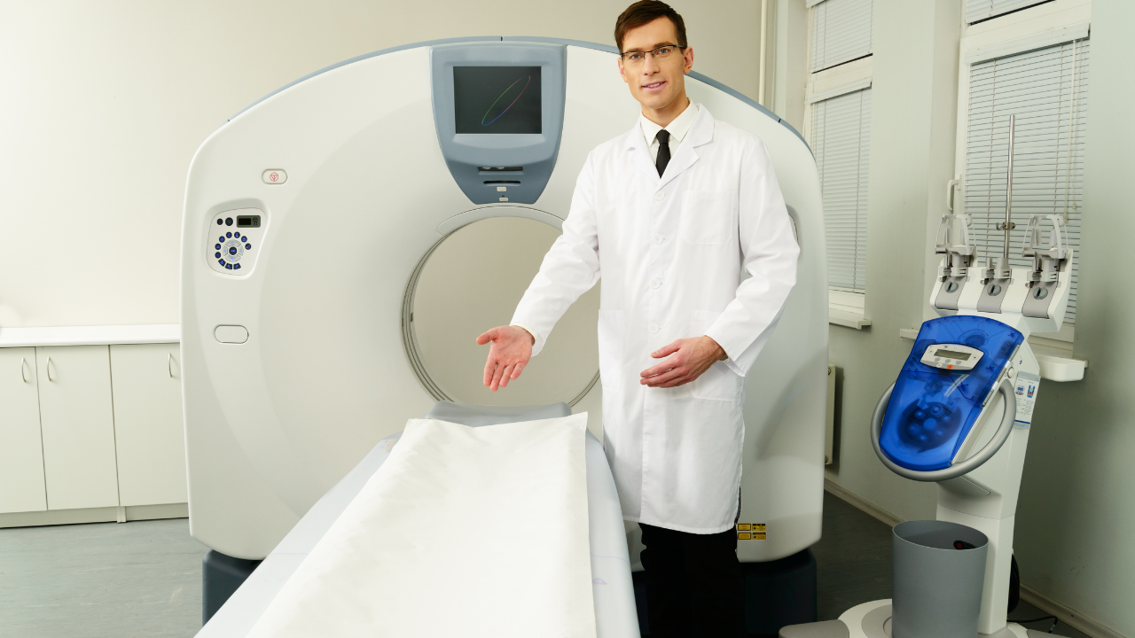
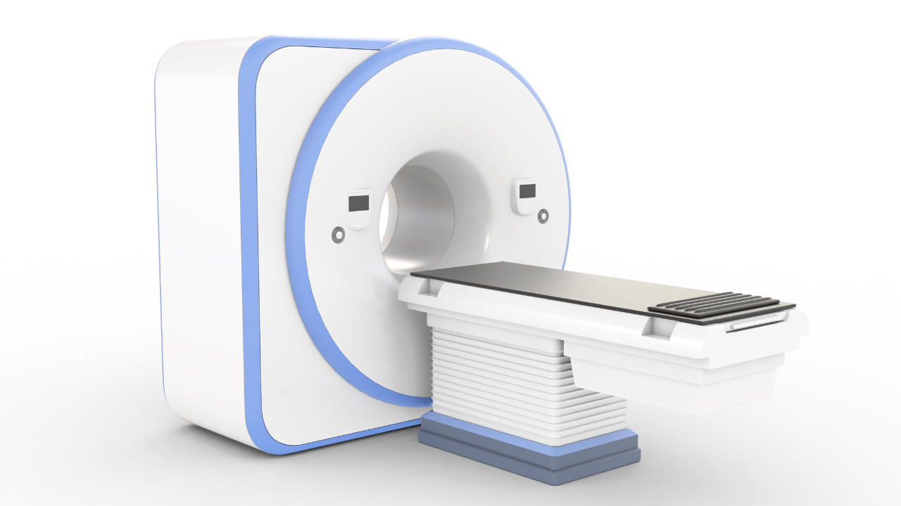
To begin with, magnetic resonance imaging is a test that uses a magnetic field and pulses of radio wave energy to make pictures of organs and structures inside the body. In many cases, MRI gives different information about structures in the body than can be seen with an X-ray, ultrasound, or computed tomography (CT) scan.
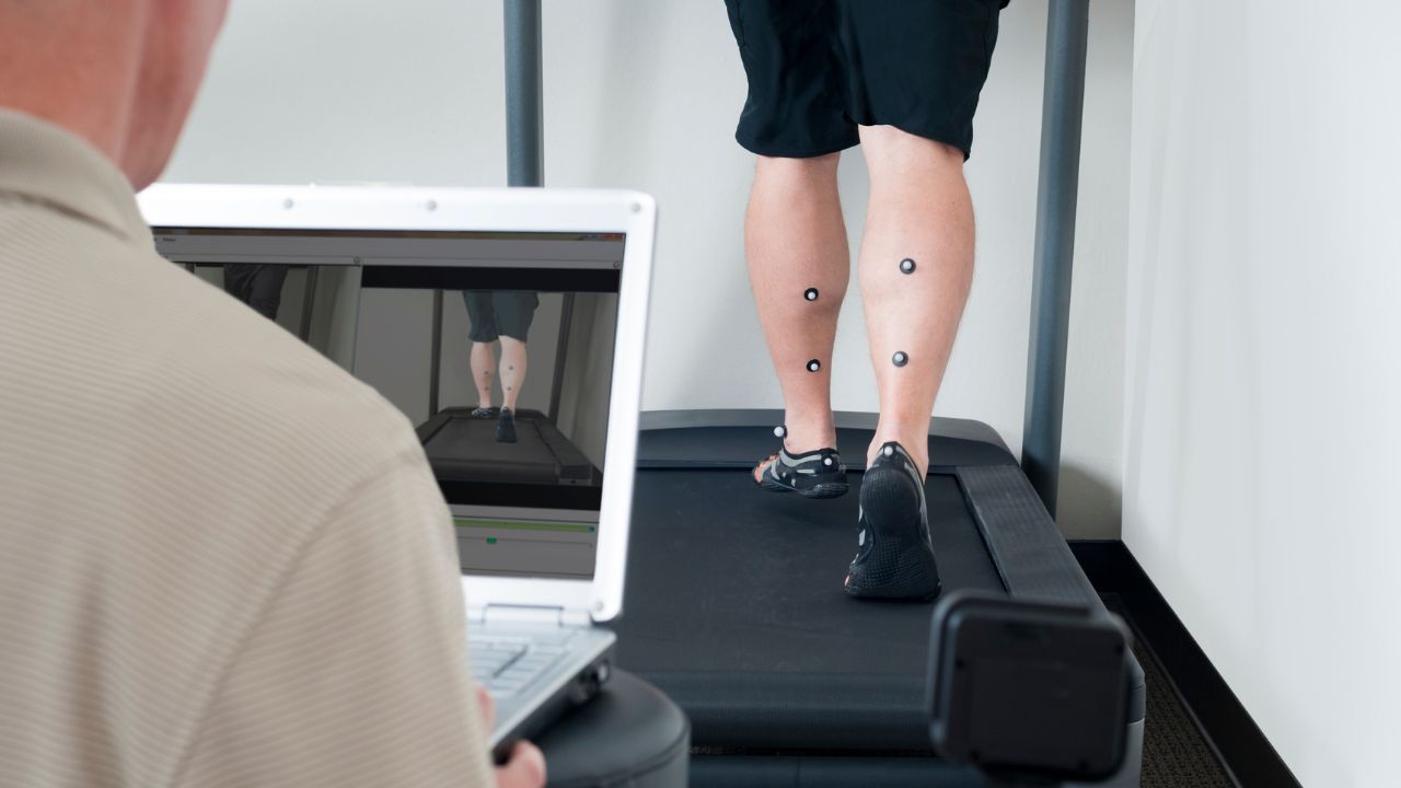
The unique soft-tissue contrast capabilities of MRI makes it a good tool for evaluating sports injuries. In many cases of knee injuries, for example, the physical finding may be hard to specify and an MRI can define the problem. MRI is greater than 90% sensitive to and specific for detecting meniscal tears and can even define tears that are best treated with primary meniscal repair.
Anterior and posterior cruciate ligament tears are also accurately defined with an MRI. MRIs may also define some bone injuries that are not diagnosed with x-rays. In complex knee injuries, MRI may detect the full spectrum of findings, which may include injuries to the menisci, cruciate and collateral ligaments and bone.
When done properly, the MRI is the only technique that can determine the degree of rotator cuff injuries as well as diagnose complete tears.
In the foot and ankle, MRIS is used to define occult bone injuries in patients with persistent pain. Tendon and ligament disruption, plantar fascitis and Achilles tendon tears can also be diagnosed with an MRI.
Thus, this technique is unique in its ability to evaluate a broad spectrum of musculoskeletal injuries non-invasively, just the type of “typical” injuries an athlete is likely to suffer from during normal play.