Blog and News

Health Care Reform
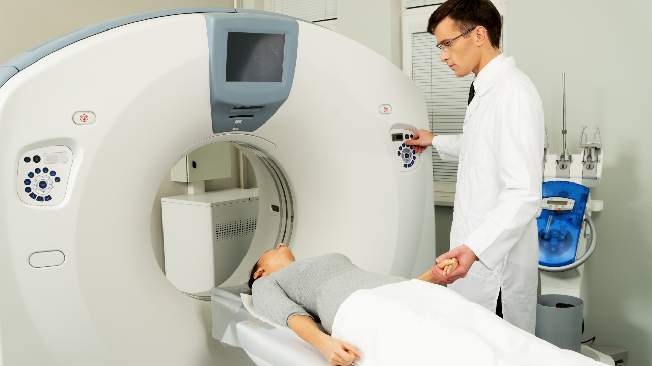
MRI – Magnetic resonance imaging — or MRI — is a test that uses a diagnostic magnetic field to pulse radio waves through the body to make pictures of organs and other internal structures. During an MRI test, the body is placed inside a special machine that contains a strong magnet (thus, these machines are usually very large). Pictures from an MRI scan are digital and can be stored on a computer for examination and diagnosis.
MRIs are performed to find problems such as tumors, internal bleeding, injuries, blood vessel diseases, and infections. In some cases, contrast material may be used during an MRI scan to show structures more clearly.
An MRI scan can be done for:
Head. MRI scans can look at the brain for tumors, aneurysms, bleeding in the brain, nerve injury and other problems, such as those related to strokes. It can also detect potential problems with the eyes, optic nerves, ears, and auditory nerves.
Chest. An MRI of the chest can examine the heart and its structures such as valves and coronary blood vessels. It can also show if the lungs are damaged. MRIs of the chest can be used to look for signs of breast or lung cancer.
Blood vessels. Using an MRI to examine blood vessels and the flow of blood through them is called a magnetic resonance angiography or MRA. This procedure can find problems with the veins and arteries, such as a blocked vessel, aneurysm or torn lining.
Abdomen and pelvis. MRIs can detect problems in the organs and other structures of the middle of the body, including the liver, gallbladder, pancreas, kidneys, and bladder. It is used to find tumors, bleeding, infection, and blockage. In men, it can be used to look at the prostate while in women, the uterus and ovaries can be examined.
Bones and joints. Arthritis, bone marrow problems, bone tumors, cartilage problems, torn ligaments and tendons, and infections can be checked with the MRI. It may also detect if a bone is broken when x-ray results are not clear.
Spine. An MRI scan can detect disc and nerves in the spine for conditions such as spinal stenosis, disc bulges and spinal tumors.
An MRI test is usually done by an MRI technologist. The pictures taken are interpreted by a radiologist. In most cases, your doctor will very likely examine the scans as well. The radiologist may discuss initial results with you right after the test. Complete results are usually ready for your doctor in one or two days.
Fox Valley Imaging provides MRI and MRA services to our patients. If your physician requests that you have an MRI, consider Fox Valley Imaging as a faster, more convenient and more economical alternative to a local hospital or other medical diagnostic services. Appointments can be made by calling 630.416.1300.
Recent Posts


Office Hours:
Monday-Friday
8:00am-5:00pm
Saturday by appointment
Services
Contact Details
Address: 1971 Gowdey Road,
Naperville, IL 60563
Phone: 630-416-1300
Fax:
630-416-1511
Email: info@foxvalleyimaging.com
© Copyright 2023 Fox Valley Imaging, Inc..