Blog and News
Bone density scans are highly accurate tools for diagnosing osteoporosis when you use well-calibrated machines and skilled operators. These scans assess mineral content in your bones through low-dose X-rays. However, several factors can lead to misdiagnosis. Operator error, such as incorrect positioning or insufficient training, can skew results. Patient movement during the scan also induces inaccuracies due to image blurring. Regular calibration of the scanning device is essential to maintain reliability. Furthermore, maintaining proper hydration is crucial as it influences the X-ray absorption and scan precision. Ensuring each of these elements is managed will enhance the diagnostic accuracy further.
Listen to the Article
Key Takeaways
- Bone density scans are generally accurate but can be affected by operator error, such as incorrect patient positioning and data entry mistakes.
- Machine calibration is crucial for ensuring scan accuracy; uncalibrated machines can lead to incorrect diagnostic outcomes.
- Patient movement during the scan can cause image blurring and shadow effects, resulting in measurement variability and possible misdiagnosis.
- Hydration levels influence scan precision, with dehydration potentially altering X-ray absorption and affecting diagnostic accuracy.
- The frequency and timing of scans are important; inappropriate intervals may not accurately reflect changes in bone density, leading to misdiagnosis.
Understanding Bone Density Scans
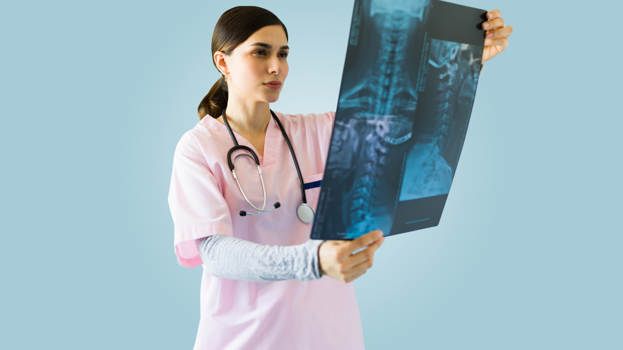
To accurately assess your risk of fractures, bone density scans, technically known as dual-energy X-ray absorptiometry (DEXA), measure the mineral content in your bones. These scans are pivotal for diagnosing conditions like osteoporosis and osteopenia, which weaken bones and increase fracture susceptibility.
A DEXA scan quantifies bone mineral density (BMD) by emitting low-dose X-rays with two distinct energy peaks, absorbed differently by bones and soft tissues. This differential absorption helps in calculating the BMD. The output, expressed in grams per square centimeter, is compared against a reference standard, typically a young healthy adult at peak bone mass. Your results are then presented as a T-score or Z-score, which indicate how many standard deviations your bone density is above or below the norm for age-matched controls or the young adult reference, respectively.
Understanding these scores is crucial. A T-score of -1.0 or above suggests normal density, between -1.0 and -2.5 indicates low bone mass (osteopenia), and lower than -2.5 points to osteoporosis, significantly raising your fracture risk. Accurate interpretation of these results can guide preventive measures or interventions, potentially mitigating severe health outcomes. Thus, these scans not only serve diagnostic purposes but are instrumental in proactive health management.
Impact of Operator Error
While bone density scans offer critical insights into bone health, operator errors can significantly impact the accuracy of these measurements. As a healthcare provider, understanding the potential errors that can arise during the scanning process is crucial. These inaccuracies not only compromise patient care but also may lead to unnecessary treatments or oversight of a necessary intervention.
Here are critical areas where operator error can influence the results:
- Incorrect Patient Positioning: Proper positioning is essential for accurate imaging. Misalignment can lead to skewed data interpretation.
- Inadequate Training on Equipment: Operators must be thoroughly trained. Incomplete understanding leads to technical mistakes.
- Failure to Account for Artifacts: Recognition and adjustment for visual obstructions or anomalies in the scan are necessary to avoid misdiagnosis.
- Inconsistent Scan Regions: Consistency in the areas scanned ensures comparability of sequential assessments. Deviation can result in inaccurate trend analysis.
- Data Entry Errors: Precise entry of patient information and scan settings is critical. Errors here can lead to incorrect analysis outcomes.
As you manage and operate these sensitive devices, it's imperative you remain vigilant about these potential pitfalls to ensure the reliability of the results and uphold your commitment to superior patient care.
Importance of Machine Calibration
Regular calibration of bone density scanning equipment is crucial to ensure the accuracy and reliability of the results you depend on for diagnosing and treating osteoporosis. Calibration involves the adjustment of the scanning device to meet required standards, using known reference points. This process ensures that the device's output remains consistent over time and across different operating conditions.
You must understand that without systematic calibration, the precision and reproducibility of bone density values can degrade, leading to potential misdiagnosis or suboptimal treatment plans. Calibration checks typically involve validating the scanner's ability to accurately measure the density of calibration phantoms —objects with known mineral densities similar to human bone. Discrepancies between the expected and measured values can indicate a need for recalibration or even repair.
Moreover, maintaining calibration logs and adherence to calibration schedules are essential practices. These records help trace the performance history of the equipment, providing invaluable data for identifying trends or deviations that might affect diagnostic decisions. As a healthcare provider committed to serving your patients effectively, it's imperative to prioritize and rigorously enforce these calibration protocols, ensuring every scan's integrity and fostering trust in the diagnostic process you support.
Effects of Patient Movement
Patient movement during a bone density scan significantly impacts the accuracy of the results, potentially leading to incorrect diagnoses. When you're engaged in conducting these scans, it's crucial to minimize any movement to ensure the precision of the data collected. Even slight shifts can alter the density measurements, which are critical for determining bone health.
Understanding the implications of patient movement can enhance your ability to serve effectively by preventing misdiagnosis. Here are key points to consider:
- Image Blurring: Movement creates a lack of sharpness in the images, reducing the clarity needed for accurate analysis.
- Shadow Effects: Sudden shifts can cause artifacts like shadowing on the scan results, which may mimic or obscure true bone structures.
- Measurement Variability: Consistent positioning is essential as variations can lead to discrepancies in bone density readings across different scans.
- Repeat Scans Necessity: Movement might necessitate redoing the scan, increasing radiation exposure and patient discomfort.
- Diagnostic Delays: Inaccuracies caused by movement can lead to further diagnostic procedures to confirm findings, delaying effective treatment.
As a professional dedicated to patient care, your attentiveness to minimizing movement can directly improve diagnostic accuracy and patient outcomes.
Analyzing Comparison Standards
To ensure the reliability of bone density scan results, it's essential to meticulously compare them against established diagnostic standards. These standards, often derived from large-scale epidemiological data, provide a benchmark that categorizes bone density levels into normal, low (osteopenia), and significantly low (osteoporosis). You'll find that precision in applying these thresholds is crucial to avoid misdiagnosis that could lead either to unnecessary treatment or to a missed opportunity for preventing further bone loss.
When you're analyzing these comparison standards, you must consider the specific calibration of the densitometry equipment used. Variations in machine calibration can lead to significant discrepancies in bone density readings. It's your responsibility to ensure that the device is consistently calibrated according to the manufacturer's specifications and validated against standardized phantoms and control groups.
Furthermore, the selection of the reference population is pivotal. The demographic characteristics of the reference group, such as age, sex, and ethnicity, should closely match those of the patient. This alignment ensures that the comparisons are clinically relevant and that the diagnostic conclusions you draw are both accurate and beneficial to patient care. By adhering to these analytical practices, you contribute significantly to the precision and reliability of bone density assessments.
Relevance of Previous Scans
Analyzing previous scans provides a historical perspective that enhances the accuracy of current bone density evaluations. When you're tasked with interpreting these crucial results, understanding the changes over time is imperative. This continuity is key in identifying subtle shifts that mightn't be apparent when viewing a single snapshot in isolation.
Here are several reasons why previous scans are indispensable:
- Baseline Comparison: Establishes a reference point, allowing for the detection of bone loss or gain over a period.
- Trend Analysis: Helps in monitoring the progression of bone density, crucial for diagnosing conditions like osteoporosis.
- Therapeutic Efficacy: Assesses the effectiveness of treatments or interventions over time.
- Technique Consistency: Ensures scan techniques and equipment haven't skewed results, offering a consistent basis for comparison.
- Patient History Contextualization: Integrates other health factors that might influence bone density, such as previous medical conditions or interventions.
Hydration Levels and Scan Accuracy
Hydration levels significantly influence the precision of bone density scans, affecting the accuracy of the results you receive. When you're dehydrated, your soft tissue composition alters, which can misleadingly suggest lower bone mineral density. This phenomenon occurs because bone density scanners, particularly those based on dual-energy X-ray absorptiometry (DXA), calculate bone density by measuring the absorption of X-ray beams by your bones. The hydration status affects the soft tissue density, which in turn influences the X-ray absorption, thereby impacting the scan's output.
To ensure optimal scan accuracy, you should maintain adequate hydration prior to undergoing a DXA scan. The scanner's calibration assumes a standard hydration level, deviations from which can lead to erroneous interpretations. For instance, a hyper-hydrated state may result in an overestimation of bone density, while dehydration typically leads to underestimation.
To serve your patients effectively, it's crucial to advise them about maintaining proper hydration before a bone density scan. This simple preparatory step can significantly enhance diagnostic accuracy, enabling more precise assessments of osteoporosis and other conditions affecting bone health. By optimizing scan conditions, you safeguard against misdiagnosis and ensure that interventions are appropriately tailored to each individual's needs.
Interval Between Scans
You must consider the optimal frequency of bone density scans to ensure accuracy without overexposure.
The timing of your scans significantly impacts the precision of the results, potentially affecting diagnosis and treatment strategies.
Adhering to recommended guidelines for repeat scans is crucial to track bone density changes accurately over time.
Optimal Scan Frequency
Determining the optimal interval between bone density scans is crucial for accurate monitoring and prevention of misdiagnosis in osteoporosis management. You'll want to balance the need for timely data with the risk of unnecessary exposure and cost.
- Patient Age: Older adults may need more frequent monitoring.
- Baseline Bone Density: Lower initial scores may warrant closer follow-up.
- Therapeutic Interventions: Adjust intervals based on treatment efficacy.
- Risk Factors: More frequent scans for those with high fracture risk.
- Technological Advances: Utilize improvements in imaging techniques to refine intervals.
Timing Impact on Accuracy
Considering the interval between bone density scans is essential, as varying the timing can significantly affect the accuracy of the results and the subsequent diagnosis.
You should understand that bone remodeling —a process where bone tissue is continuously replaced—varies over time, influencing scan results depending on the phase of remodeling when the scan is conducted.
Short intervals may not accurately reflect changes in bone density or allow for differential diagnosis of osteopenia versus osteoporosis, potentially leading to premature therapeutic interventions.
Conversely, excessively long intervals can result in missed opportunities for early intervention, escalating the risk of fractures.
Strategically timed scans are crucial for accurate monitoring and effective management of bone health, emphasizing the necessity for a balanced approach in scheduling these diagnostic evaluations.
Repeat Scan Recommendations
To optimize bone health management, it's recommended that repeat bone density scans be scheduled every two years unless specific risk factors dictate otherwise. This interval helps in accurately monitoring the progression or improvement in bone density, ensuring timely interventions.
Considerations for adjusting the frequency of bone density scans include:
- Age and Gender: Higher frequencies may be necessary for post-menopausal women or older adults who are at increased risk.
- Medication Impact: Certain medications can accelerate bone loss, necessitating closer monitoring.
- Previous Scan Results: Significant bone loss on a previous scan might warrant more frequent evaluations.
- Underlying Health Conditions: Conditions like rheumatoid arthritis or chronic renal failure affect bone density.
- Lifestyle Changes: Significant changes in diet or physical activity levels might alter bone density profiles.
Frequently Asked Questions
Can Certain Medications Interfere With Bone Density Scan Results?
Yes, certain medications can interfere with your bone density scan results, potentially leading to inaccurate readings. These include steroids, anticonvulsants, and hormone therapy drugs, which can affect bone mineral density measurements.
How Does Menopause Impact Bone Density Measurements?
Menopause significantly impacts your bone density; studies show a 20% increase in bone loss rate during this period. Hormonal changes reduce calcium absorption, crucial for maintaining strong bones, necessitating vigilant bone health monitoring.
Are Bone Density Scans Covered by Insurance?
Bone density scans might be covered by your insurance, especially if you're at risk for osteoporosis. Check your policy details or contact your provider to ensure you're eligible for this preventive diagnostic tool.
What Are the Risks of Repeated Bone Density Scans?
Radiation exposure is your primary risk with repeated bone density scans. Although minimal, it's cumulative, potentially affecting cellular structures. Carefully monitor frequency to balance diagnostic benefits against potential long-term radiation risks.
Can Dietary Choices Influence Bone Density Scan Outcomes?
Yes, your dietary choices can significantly influence bone density scan outcomes. Intake of calcium and vitamin D, particularly, affects bone health, potentially altering scan results and impacting the accuracy of osteoporosis assessments.
Recent Posts


Office Hours:
Monday-Friday
8:00am-5:00pm
Saturday by appointment
Services
Contact Details
Address: 1971 Gowdey Road,
Naperville, IL 60563
Phone: 630-416-1300
Fax:
630-416-1511
Email: info@foxvalleyimaging.com
© Copyright 2023 Fox Valley Imaging, Inc..