Blog and News
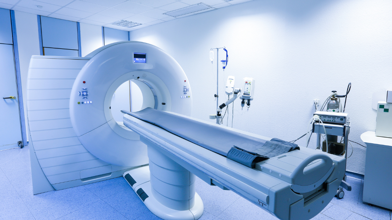
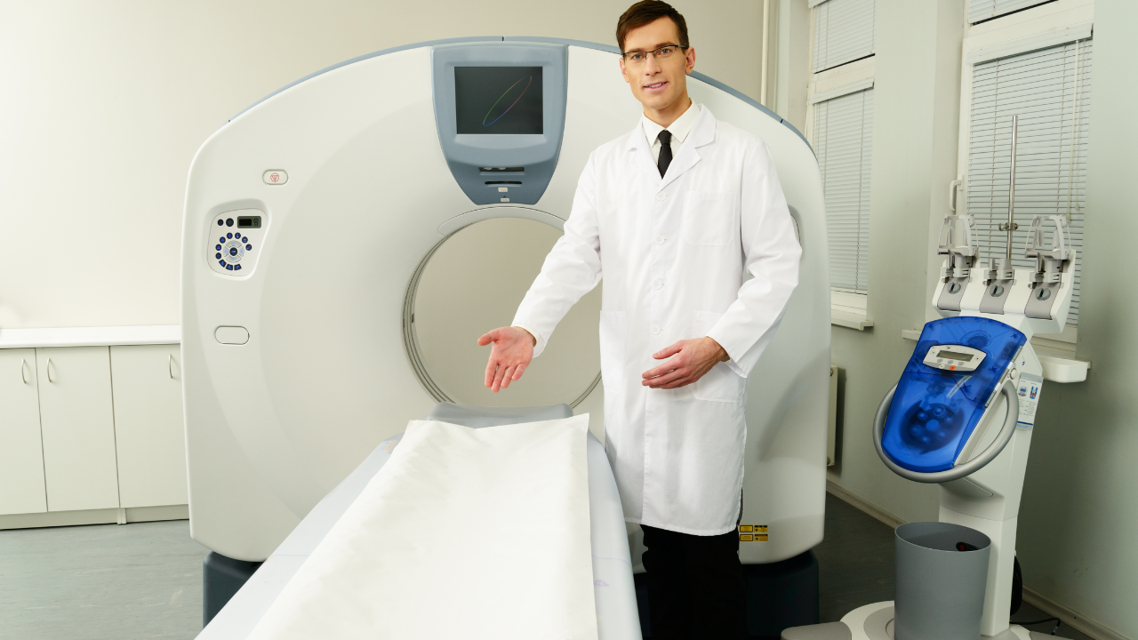
Computed tomography (CT) is fundamental in diagnosing and managing internal diseases. You'll find it invaluable for creating precise, cross-sectional images, providing clearer detail than traditional x-rays. CT scans excel in detecting and staging various cancers, from solid tumors in your chest to potentially malignant growths in your abdomen. They're also critical for assessing internal injuries, identifying blood clots, and visualizing infections with high-resolution accuracy. For ongoing disease management, CT's ability to monitor treatment efficacy and track pathological changes ensures tailored, effective interventions. Exploring further could unveil deeper insights into how these applications might directly benefit your healthcare strategies.
Listen to the Article
Key Takeaways
- Provides high-resolution images for early detection and precise localization of cancers and other internal abnormalities.
- Enables comprehensive assessment of internal injuries, often revealing issues that traditional X-rays might miss.
- Essential in identifying and monitoring blood clots, assessing risk and effectiveness of treatments.
- Facilitates detailed visualization of infections, aiding in early diagnosis and targeted treatment interventions.
- Offers crucial insights for preoperative planning and precise surgical guidance, enhancing patient safety and treatment outcomes.
Overview of Computed Tomography
What's Computed Tomography (CT), and how does it revolutionize internal disease management?
CT is a sophisticated imaging technique that utilizes x-rays and digital computer technology to produce detailed cross-sectional images of your body's internal structures. This method offers you a more precise and comprehensive view compared to traditional x-rays, allowing for enhanced diagnosis and treatment planning.
As a healthcare professional, you're aware that managing internal diseases often hinges on accurate diagnostics. CT scans provide you with high-resolution images, enabling the identification of abnormalities in organs, blood vessels, and soft tissues. By employing various contrast materials, you can further enhance the visibility of specific areas, facilitating a clearer understanding of the extent and nature of the disease.
Moreover, CT's capability to generate multiple images from different angles rapidly means you can quickly assess and respond to emergencies where time is critical. This is particularly vital in cases such as internal bleeding or infections, where rapid intervention can drastically improve outcomes.
Your role in utilizing CT technology effectively is crucial. By interpreting these detailed images, you can make informed decisions about the most appropriate treatment paths, ultimately aiding in better patient care and recovery outcomes. You're not just operating a machine; you're providing a lifeline through precise diagnostic insights.
Cancer Detection Capabilities
As a healthcare professional, you'll find that CT's advanced imaging capabilities are crucial for the early detection and precise localization of cancerous growths. Computed tomography provides high-resolution images that enable you to discern even the smallest lesions, which are often invisible on conventional radiographs. This precision is vital for staging cancer, determining the extent of tumor invasion, and planning appropriate surgical interventions.
CT scans are particularly effective for identifying and evaluating solid tumors in the chest, abdomen, and pelvis. You'll appreciate the ability to assess the size, shape, and exact location of tumors, as well as their relationship to surrounding structures. This is essential for formulating a targeted treatment strategy and predicting potential surgical challenges.
Moreover, CT's utility extends to monitoring treatment response and detecting recurrence. By comparing sequential scans, you can quantitatively measure changes in tumor size and morphology, providing a robust framework for adjusting treatment protocols and managing patient care effectively.
Understanding these capabilities, you're better equipped to make informed decisions that enhance patient outcomes. Your role in utilizing CT technology not only aids in accurate diagnosis but also significantly impacts the therapeutic approach, aligning with your commitment to serve and improve patient health.
Diagnosing Internal Injuries
When you utilize computed tomography (CT) in the evaluation of internal injuries, you're able to accurately identify hidden trauma that mightn't be apparent through conventional imaging techniques.
This modality provides a detailed assessment of organ damage, delivering crucial data that influences therapeutic decisions.
Identifying Hidden Trauma
Computed tomography (CT) excels in revealing internal injuries that traditional X-rays may overlook, offering a crucial tool for diagnosing hidden trauma. When you're faced with cases where patients exhibit ambiguous symptoms post-accident, a CT scan becomes indispensable.
It provides high-resolution images that allow for a precise evaluation of skeletal structures and soft tissues, enabling you to detect fractures, hemorrhages, or subtle dislocations that aren't visible on standard radiographs. This capability is vital; it ensures that no underlying condition goes unnoticed, thereby facilitating timely and accurate therapeutic interventions.
Your ability to identify these hidden traumas rapidly can significantly alter the course of treatment, potentially preventing long-term complications and enhancing recovery outcomes for those you're dedicated to helping.
Assessing Organ Damage
CT scans are crucial for accurately assessing organ damage and diagnosing internal injuries, providing detailed insights into the extent and nature of harm suffered. When you're analyzing conditions such as hepatic lacerations or renal contusions, the high-resolution capabilities of CT imaging allow you to visualize minute details and abnormalities that might be missed by other diagnostic tools. This precision is particularly vital in acute settings where rapid decision-making is required to optimize patient outcomes.
Identifying Blood Clots
Utilizing Computed Tomography, you can detect and analyze blood clots with high precision, crucial for timely and effective treatment. This advanced imaging technique provides cross-sectional views of the body, allowing for an in-depth assessment of vascular structures and the identification of thromboembolic events that could lead to life-threatening conditions such as strokes or pulmonary embolisms.
Computed Tomography (CT) is particularly adept at pinpointing the location and extent of a clot, distinguishing between chronic and acute thrombosis, and assessing the risk of a clot dislodging and causing further damage. Here's why the precision of CT scanning is vital:
- Early Detection: Immediate identification can mean the difference between recovery and severe complications.
- Accurate Localization: Knowing the exact position of a clot informs the approach to intervention.
- Risk Assessment: Evaluating the potential for a clot to migrate enables proactive management.
- Treatment Monitoring: CT scans assess the effectiveness of anticoagulant therapy, adjusting treatment plans as necessary.
As someone dedicated to the well-being of others, understanding these capabilities helps you advocate for the best possible diagnostic approaches, ensuring that those you serve receive timely and life-saving interventions.
Assessing Infections
In assessing infections, computed tomography provides detailed, cross-sectional images that allow for precise localization and characterization of infectious processes within the body. You'll find that CT scans are particularly valuable in visualizing areas difficult to assess through conventional imaging techniques, such as deep-seated infections in the abdomen or pelvis. The high-resolution images facilitate the identification of both the extent of an infection and the involved structures, which is crucial for effective treatment planning.
CT imaging is instrumental in distinguishing between abscess formation and other types of soft tissue infections. For instance, while you're examining a patient suspected of having an intra-abdominal abscess, CT can accurately differentiate between abscesses and other entities like cysts or tumors, based on the lesion's density and the presence of characteristic features such as an enhancing rim. This capability is vital because it directs the therapeutic approach—whether it's percutaneous drainage or more conservative management.
Moreover, in cases of osteomyelitis, CT's ability to depict cortical bone disruption and marrow involvement is superior, offering you a clear advantage in early diagnosis. This early detection enables prompt and targeted interventions, improving outcomes for those relying on your expertise to manage these complex infections effectively.
Monitoring Disease Progression
As you assess the role of computed tomography (CT) in monitoring disease progression, consider how it's pivotal in tracking treatment efficacy.
CT scans provide a detailed visual account of pathological changes over time, allowing for an analytical review of treatment impact.
You'll observe modifications in tissue structure and function, which are critical in determining the next steps in patient management.
Tracking Treatment Efficacy
Computed tomography (CT) scans are crucial for evaluating the effectiveness of treatments by providing detailed images that track disease progression or regression over time. As you use CT scans in clinical settings, it's important to recognize their value in making informed decisions on treatment modifications, enhancing patient care.
Here's how CT scans make a significant impact:
- Objective Measurement: Quantify lesion size, allowing precise assessment.
- Timely Adjustments: Detect changes early, facilitating swift treatment alterations.
- Comprehensive Overview: Visualize multiple organ systems simultaneously for holistic assessment.
- Consistent Monitoring: Provide a standardized method to compare changes over multiple time points.
Leveraging these capabilities, you can tailor treatments to individual needs, improving outcomes and demonstrating your commitment to patient-centered care.
Visualizing Pathological Changes
Building on the role of CT scans in treatment assessment, let's explore how they excel in visualizing pathological changes to monitor disease progression effectively.
You'll find that CT imaging provides high-resolution insights into anatomical anomalies and early pathological alterations that mightn't be visible with other modalities.
By analyzing cross-sectional images, you can detect subtle changes in tissue density and structure, which are crucial for diagnosing conditions like fibrosis or neoplasms.
This capability allows for precise staging of diseases, essential for tailoring treatment plans.
Moreover, periodic CT scans enable you to track the effectiveness of interventions, adjusting therapies as needed to optimize patient outcomes.
Harnessing this technology, you're equipped to make informed decisions that profoundly impact patient care.
Planning Surgical Procedures
In planning surgical procedures, utilizing computed tomography (CT) allows for precise anatomical visualization and risk assessment. As you delve into the complexities of internal diseases, it's crucial to understand that CT scans provide invaluable insights that enhance surgical outcomes. They allow you to map out a strategic approach, ensuring that every decision you make is informed and deliberate.
- Preoperative Planning: CT scans offer a three-dimensional view of the affected area, enabling you to plan the surgical route meticulously, which minimizes invasive exploration and reduces patient recovery time.
- Identification of Anomalies: You're equipped to detect any unexpected conditions that might complicate standard procedures, thereby adjusting your approach to accommodate these findings.
- Assessment of Surgical Risks: By clearly visualizing critical structures, you can predict potential complications and strategize preventive measures, thus safeguarding patient well-being.
- Enhanced Precision: The high-resolution images provided by CT technology guide you in performing precise incisions and interventions, which is particularly crucial in areas with complex anatomy.
Embracing CT in your surgical planning not only optimizes the procedure but also reflects your commitment to providing the highest standard of care. By integrating this technology, you're taking a significant step towards ensuring successful outcomes and exemplifying dedication in your service to patients.
Examining Bone Disorders
When examining bone disorders, CT scans are essential for accurately diagnosing and assessing the extent of skeletal abnormalities. You'll find their high-resolution images allow for a detailed visualization of bone density, structure, and integrity, which is crucial in identifying conditions such as osteoporosis, fractures, or malignancies like osteosarcoma. The cross-sectional imaging capability of CT is particularly useful in delineating the architecture of complex bone structures and can be pivotal in planning surgical interventions.
Moreover, CT technology excels in quantifying bone mineral density, which aids in the evaluation of diseases like osteopenia. This precise measurement can guide you in making informed decisions about medication or lifestyle adjustments necessary for managing the disease effectively. In cases of traumatic injuries, CT scans provide a clear picture of the extent of damage, helping you to strategize the most appropriate surgical or non-surgical treatments tailored to individual patient needs.
In your service to patients suffering from bone disorders, utilizing CT imaging can facilitate a more accurate diagnosis, monitor progression or healing, and ultimately guide therapeutic strategies. This ensures that your interventions aren't only timely but also specifically adapted to the needs of each patient, enhancing both care outcomes and patient satisfaction.
Organ Examination Accuracy
Just as CT scans significantly enhance bone disorder diagnostics, they also provide unparalleled accuracy in examining soft tissue organs such as the liver, pancreas, and kidneys. You'll appreciate how this imaging tool plays a pivotal role in diagnosing and managing various conditions, enabling you to serve patients with precision and empathy.
- Liver: CT scans detect early signs of liver disease, including fatty liver, cirrhosis, and tumors. This early detection is crucial for timely intervention, which can significantly change a patient's prognosis.
- Pancreas: By capturing detailed images, CT technology helps identify pancreatic cysts and cancers, which are often elusive on other imaging modalities. Early diagnosis can be life-saving.
- Kidneys: Using CT scans, you can detect structural abnormalities, stones, and masses in the kidneys. This facilitates early management strategies, reducing the risk of long-term complications.
- Adrenal Glands: CT scans are instrumental in identifying adrenal tumors and hyperplasias, which, if left unchecked, could lead to severe hormonal imbalances.
As you utilize CT imaging, you're not just operating a machine; you're extending a lifeline to those in dire need of accurate diagnosis and compassionate care.
Frequently Asked Questions
What Are the Common Side Effects of CT Scans?
You may experience mild side effects from CT scans, such as nausea, hives, or a metallic taste. Rarely, serious reactions like kidney issues from contrast dye occur, demanding vigilant post-scan monitoring for safety.
How Long Does a CT Scan Typically Take?
A CT scan typically takes 10 to 30 minutes, depending on the complexity of the area being examined. You'll lie still as the scanner captures detailed images crucial for diagnosing internal conditions.
Can Pregnant Women Safely Undergo a CT Scan?
If you're pregnant, CT scans are generally avoided due to radiation risks to the fetus. However, in urgent cases, such as suspected pulmonary embolism, it might be necessary with appropriate shielding and dose adjustments.
What Is the Cost Range for a CT Scan?
The cost of a CT scan varies widely, typically ranging from $300 to $6,000, depending on factors such as location, facility type, and whether contrast materials are used during the procedure.
Are There Alternative Imaging Techniques to CT Scans?
Yes, alternatives to CT scans include MRI and ultrasound, offering detailed glimpses without radiation. These techniques provide diverse insights, crucial for those dedicated to patient care, tailoring diagnosis to individual health needs.
Recent Posts


Office Hours:
Monday-Friday
8:00am-5:00pm
Saturday by appointment
Services
Contact Details
Address: 1971 Gowdey Road,
Naperville, IL 60563
Phone: 630-416-1300
Fax:
630-416-1511
Email: info@foxvalleyimaging.com
© Copyright 2023 Fox Valley Imaging, Inc..