Blog and News

Bone density scanning, especially DEXA, accurately predicts your risk of osteoporosis by measuring your bone mineral density (BMD). This scan compares your T-score against healthy young adults and your Z-score with others in your demographic group. If you have a T-score of -2.5 or lower, it indicates osteoporosis, putting you at a higher risk for fractures. Regular scans can track changes in your bone density, allowing timely interventions. These proactive measures can reduce your fracture risk significantly. Understanding these results equips you with the information needed to make informed decisions about preventative strategies and treatments. Explore further to understand how these insights apply specifically to you.
Listen to the Article
Key Takeaways
- Bone density scanning measures BMD, crucial for diagnosing osteoporosis and assessing fracture risk.
- T-scores from scans compare patient bone density to a healthy 30-year-old reference, predicting osteoporosis.
- Regular screenings track BMD changes, allowing timely adjustments in treatment to prevent further bone loss.
- FRAX algorithm uses scan results to evaluate individual fracture risk, guiding intervention strategies.
- Early detection through scans enables interventions such as dietary and exercise modifications to manage osteoporosis effectively.
Understanding Osteoporosis
Osteoporosis, a condition characterized by weakened bones and an increased risk of fractures, affects millions globally, making it crucial to understand its underlying mechanisms and risk factors. You'll find that the disease primarily stems from an imbalance between bone resorption and bone formation. As you age, bone mass and strength decrease, a process exacerbated by hormonal changes, particularly in postmenopausal women due to reduced estrogen levels.
Research indicates that several factors contribute to osteoporosis risk. Genetics play a significant role; a family history of osteoporosis can increase your predisposition. Dietary factors are also critical, especially inadequate intake of calcium and vitamin D, which are essential for bone health. Furthermore, lifestyle choices such as smoking and excessive alcohol consumption have been linked to bone density loss.
Physical inactivity further elevates the risk; regular weight-bearing exercises help maintain and improve bone density. Additionally, certain medications and medical conditions can also predispose individuals to osteoporosis, making it essential for healthcare providers to assess these risks when treating patients.
Understanding these factors allows you to take proactive steps toward prevention and management, emphasizing the importance of early intervention and tailored healthcare strategies to mitigate the impact of this debilitating disease.
What Is Bone Density Scanning?
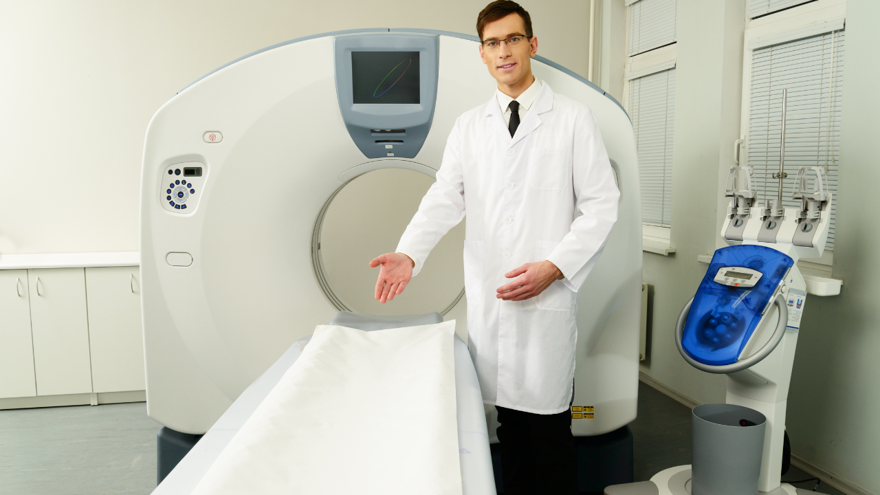
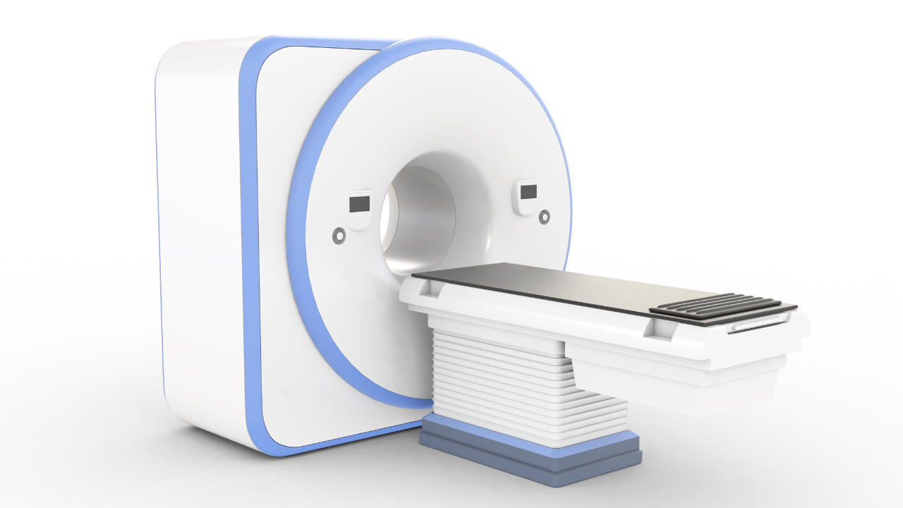
Bone density scanning, also known as dual-energy X-ray absorptiometry (DXA), precisely measures the mineral content of your bones to assess fracture risk and diagnose osteoporosis. This non-invasive test provides critical data that helps healthcare professionals manage and mitigate the debilitating impacts of osteoporosis, particularly in populations at increased risk, such as post-menopausal women and older adults.
When you undergo a bone density scan, several key aspects are evaluated:
- Bone Mineral Density (BMD): This is the primary measurement taken during a DXA scan. It quantifies the calcium and other minerals packed into a segment of your bone, providing a snapshot of your skeletal strength.
- T-score: Your BMD results are compared against a young healthy reference population, typically at peak bone mass. This comparison yields your T-score, which is crucial in diagnosing osteoporosis.
- Z-score: This score compares your BMD with what's typically expected for someone of your age, sex, and body size. It helps determine if your bone loss is within normal ranges or suggestive of a medical condition.
Understanding these outputs empowers you and your healthcare provider to make informed decisions about interventions that can slow bone loss, improve bone density, and significantly reduce the risk of fractures.
The Role of DEXA Scans
Understanding the outputs from bone density scanning, DEXA scans serve as a fundamental tool in both diagnosing and monitoring the progression of osteoporosis. DEXA, or Dual-Energy X-ray Absorptiometry, measures bone mineral density (BMD) with high precision, offering critical insights into bone health. This technique utilizes low-dose X-rays at two different energy levels to accurately differentiate between bone and soft tissue, providing a detailed image of bone integrity.
The quantitative data you obtain from a DEXA scan helps in identifying decreases in bone density, thus facilitating early intervention strategies. Regular DEXA scans are paramount in assessing the effectiveness of osteoporosis treatment, allowing adjustments based on empirical evidence. The procedure is non-invasive, quick, and the radiation exposure is minimal, making it safe for frequent monitoring.
In clinical practice, the use of DEXA scans has been instrumental in reducing fracture risks by enabling timely preventive measures in individuals at high risk. As you strive to protect and improve patient health, integrating DEXA scans into routine assessments can be a proactive approach to mitigating severe osteoporotic complications. This tool not only aids in precise diagnosis but also enhances your ability to guide patients towards maintaining stronger bones and a healthier life.
Interpreting T-Scores and Z-Scores
When interpreting DEXA scan results, it's crucial to understand T-scores and Z-scores, which provide quantitative measures of bone density compared to established norms. T-scores compare your bone density to a healthy 30-year-old adult's average density, which is considered the peak bone density age. Z-scores, on the other hand, compare your bone density to what's typical for someone of your age, gender, and size.
Understanding these scores helps in identifying varying degrees of bone density, giving insights into your bone health:
- T-score of -1.0 or above: Your bone density is considered normal.
- T-score between -1.0 and -2.5: This indicates osteopenia, a condition where bone density is lower than normal and may lead to osteoporosis.
- T-score of -2.5 or lower: This score suggests a diagnosis of osteoporosis.
The Z-score is particularly useful if you're under 50 years old, as it accounts for expected bone density variations based on demographic factors. A significantly lower Z-score than expected might prompt further medical evaluation for secondary causes affecting bone loss.
Accurate interpretation of these scores allows you to take proactive steps towards managing your bone health, ensuring you can continue to serve others effectively without the personal risk of bone injuries.
Assessing Fracture Risk
As you assess fracture risk, it's crucial to identify individuals with a high probability of osteoporotic fractures based on their bone density scores and other clinical factors. Research shows that integrating these scores with tools like FRAX provides a robust prediction of fracture risk, guiding the implementation of targeted preventative strategies.
Identifying High-Risk Individuals
To effectively assess fracture risk, clinicians use bone density scanning to identify individuals at high risk for osteoporosis. This diagnostic tool measures bone mineral density (BMD), providing crucial data that helps you understand your patients' bone health. By quantifying BMD, you can pinpoint those who are more susceptible to fractures.
Key factors contributing to high fracture risk include:
- Age: The risk increases significantly with age, particularly in postmenopausal women.
- Family History: A familial predisposition to osteoporosis can elevate an individual's risk.
- Previous Fractures: Patients with a history of fractures are at a higher risk of subsequent breaks.
Utilizing this information allows you to tailor interventions and monitor those who need the most vigilant care.
Enhancing Preventative Strategies
Enhancing preventative strategies involves rigorously assessing fracture risk through evidence-based methods and updated clinical guidelines. You'll utilize tools like the FRAX algorithm, which integrates bone density data with individual risk factors —age, gender, smoking status, and previous fractures—to calculate the probability of breaking a bone within the next ten years.
This approach allows you to tailor interventions more precisely, prioritizing individuals at higher risk. By incorporating these quantitative assessments, you're not just treating osteoporosis, you're actively preventing its most severe consequences. Moreover, ongoing research continually refines these tools, enhancing their predictive accuracy.
As a dedicated healthcare provider, your commitment to applying these advanced techniques ensures better preventative care and a significant reduction in osteoporotic fractures among your patients.
Monitoring Bone Loss
Monitoring bone loss is crucial for managing osteoporosis. You'll find that early detection techniques are key to preventing significant density reductions.
Regular screening benefits include timely interventions that can halt or reverse bone degradation.
Advanced imaging technologies provide precise data, allowing for targeted treatment plans and improved outcomes.
Early Detection Techniques
Detecting bone loss early through advanced scanning technologies significantly increases the likelihood of preventing osteoporosis-related fractures. Utilizing Dual-energy X-ray Absorptiometry (DXA), you can accurately measure bone mineral density (BMD). This approach is crucial, as early changes in BMD can be subtle yet predict substantial risk.
- DXA Scans: Provide quantitative data on bone density, allowing for targeted interventions.
- Quantitative Ultrasound (QUS): Offers a portable alternative to DXA, assessing bone quality and strength.
- T-score Analysis: Compares your bone density with a healthy young adult's baseline, quantifying deviation and risk.
These techniques empower healthcare providers to implement preventative strategies effectively and tailor treatments to individual needs, significantly enhancing patient care and outcomes.
Regular Screening Benefits
By incorporating regular screening into your healthcare routine, you can track changes in bone density and adjust treatments proactively to mitigate the risk of osteoporosis. Regular monitoring through bone density scans, such as Dual-Energy X-ray Absorptiometry (DXA), provides quantifiable data, allowing healthcare providers to detect even subtle losses in bone density earlier than clinical symptoms might suggest.
This early intervention is crucial, as it supports the modification of treatment plans, potentially incorporating dietary adjustments, tailored exercise regimens, and pharmacotherapy to enhance bone strength and reduce fracture risk. Studies indicate that patients undergoing biennial screenings show a significant reduction in osteoporotic fractures compared to those screened less frequently, highlighting the importance of consistent monitoring in effective osteoporosis management.
Advanced Imaging Technologies
Advanced imaging technologies, such as High-Resolution Peripheral Quantitative Computed Tomography (HR-pQCT), now enable you to observe minute changes in bone texture and strength, providing critical data for early osteoporosis intervention. This technology offers unprecedented precision in detecting and quantifying even the smallest alterations in bone density and microarchitecture that traditional scans might miss.
Benefits of HR-pQCT include:
- Enhanced Precision: Detects micro-architectural deterioration that DXA scans cannot.
- Targeted Treatment: Allows for tailored interventions based on specific site and type of bone loss.
- Progress Monitoring: Tracks the effectiveness of treatment over time with high accuracy.
Importance of Baseline Measurements
Establishing baseline bone density measurements is crucial for accurately tracking the progression of osteoporosis in patients. You'll find that initial screenings provide a reference point, helping you and healthcare providers to monitor changes over time. This initial measurement is typically conducted using dual-energy X-ray absorptiometry (DEXA), which quantifies bone mineral density (BMD).
The data from your baseline DEXA scan are used to calculate your T-score, which compares your bone density to a healthy 30-year-old's average. A T-score of -1.0 or above is considered normal, while scores between -1.0 and -2.5 indicate low bone mass, or osteopenia, and scores below -2.5 confirm osteoporosis.
Subsequent scans, recommended every two to five years depending on initial results and risk factors, are essential for assessing the rate of bone loss. This is pivotal in tailoring your treatment and prevention strategies. For instance, if your follow-up scans show significant bone density reduction, your healthcare provider might adjust your treatment regimen, perhaps increasing calcium and vitamin D intake or prescribing specific osteoporosis medications.
Considering Radiation Exposure
When considering bone density scanning, it's important to evaluate the radiation exposure associated with DEXA tests. Dual-energy X-ray absorptiometry (DEXA) scans are the gold standard for measuring bone mineral density, crucial for diagnosing osteoporosis. However, understanding the balance between diagnostic benefits and the risks of radiation exposure is essential for making informed healthcare decisions.
DEXA scans expose you to a low dose of ionizing radiation. To put this into perspective, the radiation dose from a standard DEXA scan is about 0.001 millisieverts (mSv), which is equivalent to the natural background radiation received over about one day in the U.S. This is substantially lower than other imaging tests, such as a standard chest X-ray, which delivers about 0.1 mSv.
To emphasize the context of radiation in DEXA scans:
- Comparative Safety: DEXA scans are among the lowest in terms of radiation exposure compared to other diagnostic radiological procedures.
- Cumulative Dose: For most patients, the cumulative radiation dose from regular DEXA scans is minimal.
- Risk-Benefit Analysis: The potential health risks from radiation are significantly outweighed by the benefits of detecting osteoporosis early.
Understanding these points can help you make more informed decisions about your healthcare, prioritizing your safety while effectively managing the risk of osteoporosis.
Frequency of Scans
Determining the optimal frequency of DEXA scans involves assessing individual risk factors and bone loss rates to prevent overexposure while ensuring effective monitoring of osteoporosis progression. For you, this means considering personalized factors such as age, gender, family history, and previous fracture incidents.
Guidelines suggest that initial screening mightn't need repetition for about 10 to 15 years if the results are normal and no risk factors emerge. However, if you're at a higher risk or your initial scan shows bone density loss, more frequent monitoring is crucial. Typically, healthcare professionals recommend a follow-up scan every two years to assess the effectiveness of osteoporosis treatment and adjust if necessary.
Data from large cohort studies indicate that the risk of fracture correlates strongly with bone density scores. Monitoring changes over time allows for timely interventions, potentially reducing fracture risk significantly. As someone serving others, it's your responsibility to ensure that patients are neither under-monitored, risking unnoticed progression, nor over-monitored, leading to unnecessary radiation exposure and healthcare costs.
Frequently Asked Questions
Can Children Undergo Bone Density Scanning?
Yes, children can undergo bone density scanning, especially if they're at risk for conditions affecting bone strength. It's a crucial step in safeguarding their skeletal health as they grow and develop.
Are There Alternatives to DEXA for Bone Density Testing?
Yes, alternatives to DEXA for bone density testing include quantitative ultrasound and CT-based absorptiometry. These methods provide valuable data, helping you effectively serve those at risk for bone density issues.
How Does Menopause Impact Bone Density Results?
During menopause, your estrogen levels drop significantly, which can lead to decreased bone density. Regular screenings help identify this change, empowering you to take preventive actions and manage your bone health effectively.
Can Lifestyle Changes Improve Bone Density Scores?
Yes, you can improve your bone density scores through lifestyle changes like increased physical activity, calcium-rich diets, and vitamin D supplementation, all supported by data showing significant benefits in bone health maintenance and enhancement.
What Medications Affect Bone Density Scan Accuracy?
Certain medications you're taking can skew your bone density scan results. Drugs like steroids and proton pump inhibitors are culprits. Understanding these influences helps ensure your scans accurately reflect your bone health status.
Recent Posts


Office Hours:
Monday-Friday
8:00am-5:00pm
Saturday by appointment
Services
Contact Details
Address: 1971 Gowdey Road,
Naperville, IL 60563
Phone: 630-416-1300
Fax:
630-416-1511
Email: info@foxvalleyimaging.com
© Copyright 2023 Fox Valley Imaging, Inc..