Blog and News
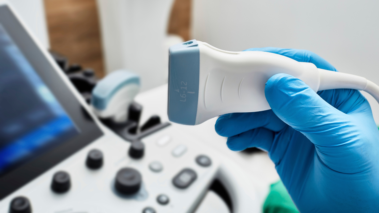
Using Doppler ultrasound techniques can greatly optimize vascular diagnostics. Choose the right Doppler mode—continuous wave for high-velocity flow, pulsed wave for precise localization, and color Doppler for visual flow direction. Apply ultrasound gel thoroughly to avoid air pockets and ensure patient comfort. Focus on major blood vessels and pinpoint abnormalities. Position the probe parallel to the vessel to minimize angle error and maintain consistent pressure. Listen for blood flow sounds to detect stenosis or occlusions. By adjusting gain settings and accurately interpreting waveforms, you can provide precise assessments. Delve deeper to explore these techniques' full potential in enhancing patient outcomes.
Key Takeaways
- Continuous Wave Doppler measures high-velocity blood flow, identifying significant stenosis or occlusions.
- Pulsed Wave Doppler provides precise localization of blood flow abnormalities, enhancing targeted diagnostics.
- Color Doppler visualizes flow direction and turbulence, aiding in the detection of vascular disorders.
- Proper application of ultrasound gel ensures optimal transmission, preventing vasoconstriction and improving diagnostic accuracy.
- Correct probe positioning minimizes angle error and compression, ensuring high-resolution imaging and accurate assessment.
Choosing the Right Doppler Mode
Selecting the appropriate Doppler mode is crucial for accurately diagnosing various vascular conditions and ensuring optimal patient outcomes.
You should choose between Continuous Wave Doppler for high-velocity blood flow, Pulsed Wave Doppler for precise localization, or Color Doppler for visualizing flow direction.
Each mode provides unique insights, enabling you to tailor your approach for comprehensive vascular assessment and improved care.
Applying Gel to the Skin
Applying an adequate amount of ultrasound gel to the patient's skin is essential to ensure optimal transmission of sound waves and clear imaging results. Use a liberal amount to cover the area thoroughly, eliminating air pockets that could interfere with the scan.
The gel should be at room temperature to maximize patient comfort and prevent vasoconstriction, which could impact diagnostic accuracy.
Selecting the Area of Interest
Once the gel is adequately applied, you should identify the exact anatomical location where the Doppler ultrasound will provide the most diagnostic value.
Focus on major blood vessels, such as the carotid artery or femoral vein.
Pinpoint areas with suspected vascular abnormalities, like stenosis or thrombosis.
Accurate selection enhances diagnostic outcomes, ensuring you provide the best possible care for your patients.
Positioning the Probe Correctly
Properly positioning the probe is crucial for obtaining accurate and meaningful Doppler ultrasound measurements in vascular diagnostics. Ensure the probe is aligned parallel to the vessel to minimize angle error.
Maintain consistent pressure to avoid compressing the vessel, which could alter flow readings.
Regularly adjust the probe orientation to follow the vessel's course, ensuring optimal image resolution and reliable data for patient care.
Listening for Blood Flow Sounds
When you listen for blood flow sounds, focus on identifying any irregularities that could indicate abnormal blood flow. This practice significantly enhances diagnostic accuracy by allowing you to detect conditions like stenosis or occlusions.
Ensure you meticulously interpret the auditory signals to provide precise and effective patient care.
Identifying Abnormal Blood Flow
By meticulously listening for blood flow sounds with a Doppler ultrasound, you can accurately identify abnormal vascular conditions such as stenosis or occlusions.
Recognizing turbulent or disrupted flow patterns is crucial. These atypical sounds indicate narrowing or blockage, requiring prompt intervention.
Detailed auditory analysis allows you to pinpoint issues, ensuring timely, patient-focused care and improving vascular health outcomes through early detection and management.
Enhancing Diagnostic Accuracy
How can you enhance diagnostic accuracy when listening for blood flow sounds with Doppler ultrasound?
Utilize high-frequency transducers to capture detailed waveforms.
Focus on angle correction to ensure precise velocity measurements.
Adjust gain settings to optimize signal-to-noise ratio.
Train thoroughly to differentiate between normal and pathological flow patterns.
Your meticulous approach ensures patients receive accurate, life-saving diagnostics.
Adjusting the Gain for Clarity
Fine-tuning the gain on your Doppler ultrasound device enhances the visualization of vascular structures, ensuring accurate diagnostic results.
Adjusting the gain helps minimize noise, providing a clearer image of blood flow and vessel walls.
Start with a lower gain and gradually increase it until the vessels and flow are distinct without excessive background artifacts, improving the quality and reliability of your vascular assessments.
Interpreting the Waveform
Interpreting the waveform in Doppler ultrasound involves analyzing the shape, amplitude, and frequency of the signal to assess the hemodynamics and identify potential vascular abnormalities.
You should focus on:
- Peak systolic velocity to detect stenosis.
- End-diastolic velocity to evaluate downstream resistance.
- Waveform shape to differentiate between normal and abnormal flow patterns.
Accurate interpretation is vital for diagnosing conditions and guiding patient care.
Measuring Systolic and Diastolic Velocities
When measuring systolic and diastolic velocities, you need to ensure velocity measurement accuracy to provide reliable diagnostic data.
Accurate measurements are crucial for gaining clinical insights into blood flow dynamics and identifying potential vascular issues.
These diagnostic applications help you tailor treatment plans to the specific needs of each patient.
Velocity Measurement Accuracy
Accurately measuring systolic and diastolic velocities with Doppler ultrasound is crucial for effective vascular diagnostics and patient management.
You need to ensure precision by focusing on:
- Angle Correction: Align the ultrasound beam with blood flow direction.
- Sample Volume Placement: Position it centrally within the vessel.
- Spectral Analysis: Utilize advanced software for detailed waveform analysis.
These steps enhance diagnostic accuracy, leading to better patient outcomes.
Clinical Relevance Insights
Understanding systolic and diastolic velocities through Doppler ultrasound provides critical insights into a patient's vascular health, directly impacting diagnostic decisions and treatment plans.
Diagnostic Applications
By harnessing Doppler ultrasound to measure systolic and diastolic velocities, you can precisely evaluate blood flow characteristics and identify vascular abnormalities with greater accuracy. This technique allows for:
- Early detection of arterial stenosis
- Detailed assessment of venous insufficiency
- Monitoring of post-surgical vascular grafts
Your dedication to utilizing these advanced diagnostic tools ensures better patient outcomes and enhances the quality of vascular care.
Documenting the Findings
When documenting the findings from a Doppler ultrasound, ensure you include detailed descriptions of blood flow characteristics, vessel patency, and any abnormalities detected.
Highlight the velocity, turbulence, and direction of blood flow. Note any stenosis, occlusions, or aneurysms.
Use precise measurements and clear terminology to aid in diagnosis and treatment planning. Your thorough documentation can significantly impact patient outcomes and facilitate effective communication with the healthcare team.
Frequently Asked Questions
How Does Doppler Ultrasound Differ From Traditional Ultrasound?
Doppler ultrasound provides real-time blood flow information, unlike traditional ultrasound which captures static images. You'll use Doppler to assess vascular conditions more accurately, enhancing patient care by identifying abnormalities like blockages or blood clots promptly.
What Are the Common Indications for a Doppler Ultrasound?
Oh, you didn't want to know about detecting blood flow issues, right? Common indications include deep vein thrombosis, carotid artery stenosis, and peripheral artery disease. You're in it to serve, after all, ensuring accurate, patient-focused diagnostics.
Are There Any Risks or Side Effects Associated With Doppler Ultrasound?
You won't experience significant risks or side effects with Doppler ultrasound. It's non-invasive and doesn't use ionizing radiation. Some patients might feel slight discomfort from the transducer's pressure, but it's generally well-tolerated.
Can Doppler Ultrasound Detect Blood Clots?
Absolutely, Doppler ultrasound can detect blood clots by measuring blood flow abnormalities. You'll find it invaluable in diagnosing deep vein thrombosis. It's non-invasive, ensuring patient comfort while providing crucial diagnostic information for effective treatment planning.
How Long Does a Typical Doppler Ultrasound Procedure Take?
A typical Doppler ultrasound procedure usually takes about 30 to 60 minutes. You'll have a technologist apply gel and move a transducer over your skin to assess blood flow, ensuring accurate vascular diagnostics for optimal patient care.
Conclusion
By mastering Doppler ultrasound techniques, you'll significantly enhance vascular diagnostics. Imagine a patient with unexplained leg pain. Using precise probe positioning and waveform interpretation, you identify a deep vein thrombosis early.
This timely diagnosis not only alleviates the patient's discomfort but potentially saves their life. Documenting such critical findings ensures continuity of care. Your expertise and attention to detail make a profound difference in patient outcomes, turning technology into life-saving action.
Recent Posts


Office Hours:
Monday-Friday
8:00am-5:00pm
Saturday by appointment
Services
Contact Details
Address: 1971 Gowdey Road,
Naperville, IL 60563
Phone: 630-416-1300
Fax:
630-416-1511
Email: info@foxvalleyimaging.com
© Copyright 2023 Fox Valley Imaging, Inc..