Blog and News
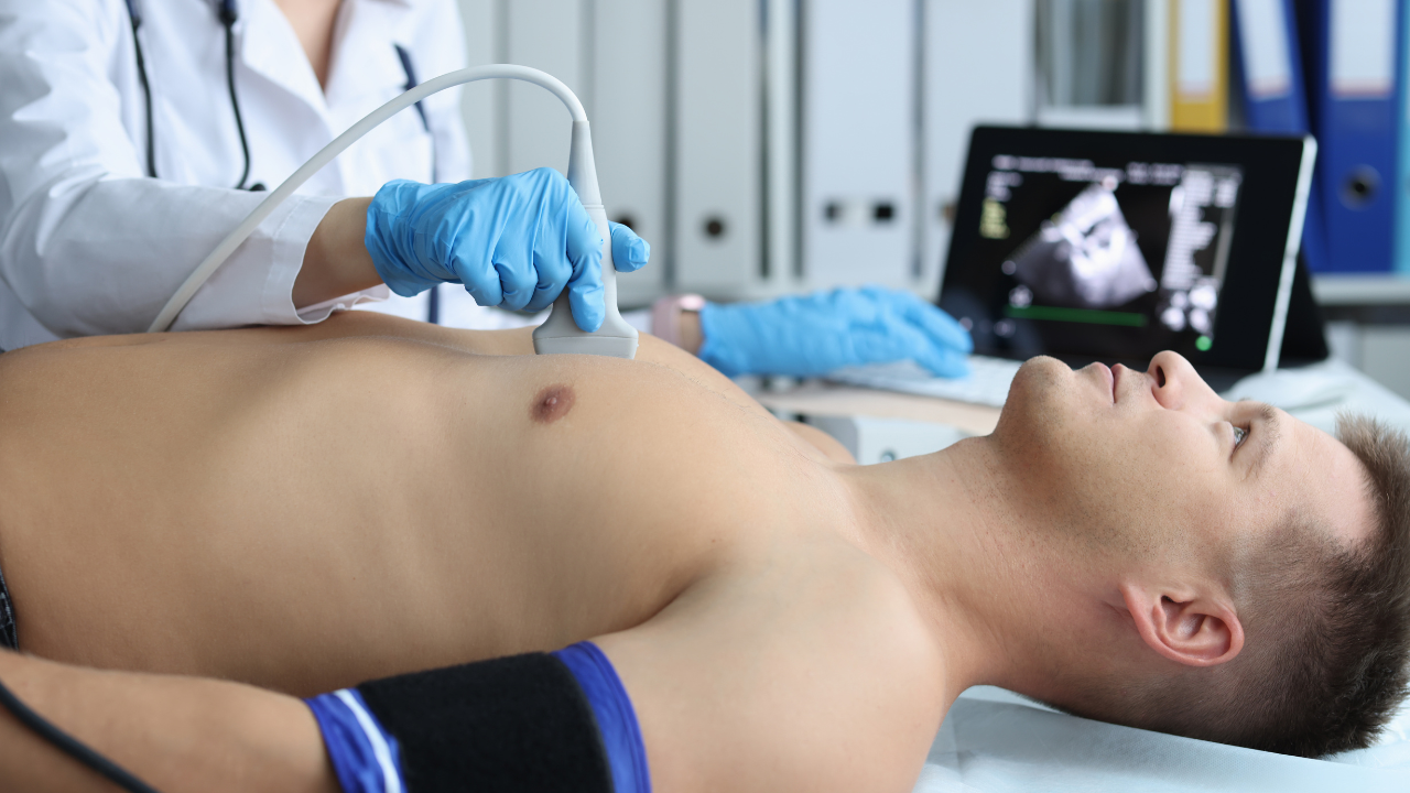
Echocardiography allows you to see detailed images of your heart without needing surgery by using high-frequency sound waves. A device called a transducer emits these waves, which bounce off your heart tissues and return as echoes. These echoes are then captured and converted into real-time, dynamic images of your heart in action. This method provides critical information on heart structure, function, and any abnormalities, aiding in accurate diagnosis and treatment planning. It's non-invasive, avoids the risks associated with surgical procedures, and can be performed quickly, even at your bedside. As you explore further, you'll discover even more about the pivotal role echocardiography plays in modern cardiac care.
Listen to the Article
Key Takeaways
- Echocardiography uses high-frequency sound waves to create detailed images of the heart's structure and function.
- It is a non-invasive method, avoiding incisions and general anesthesia, enhancing patient safety and comfort.
- Various types, including TEE and TTE, allow for comprehensive viewing of different heart areas without surgical intervention.
- Real-time imaging capabilities enable dynamic assessment of the heart under different physiological conditions.
- Utilizes ultrasound technology, where sound waves are reflected off heart tissues to form images, eliminating the need for surgical exposure.
Understanding Echocardiography Basics
Echocardiography, often referred to as an 'echo,' uses sound waves to create detailed images of your heart's structure and function. This technique is non-invasive and provides critical data that can guide diagnoses, monitor therapy, and inform surgical decisions, especially beneficial in settings committed to serving and healing others.
You'll find that during an echocardiogram, a device called a transducer is placed on your chest. It emits high-frequency sound waves that bounce off your heart structures, sending echoes back to the transducer. These echoes are converted into live, moving images of your heart, visible on a monitor. This process allows healthcare providers to see your heart beating and pumping blood, granting them the ability to assess the size and shape of your heart, the functioning of its chambers and valves, and the presence of any abnormalities.
This imaging tool is indispensable in diagnosing a range of heart conditions, from infections and disease to defects you've had since birth. By providing a clear picture of heart health, echocardiography can help medical professionals develop a targeted treatment plan that serves the patient's specific needs, enhancing both the quality and effectiveness of care.
Types of Echocardiography Explained
Several types of echocardiograms are available, each tailored to uncover specific aspects of heart function and structure. The most common type you'll encounter is the transthoracic echocardiogram (TTE). It's non-invasive and provides a comprehensive view of the heart using a transducer placed on the chest wall. It allows for the evaluation of the heart chambers, valves, and the pericardium.
Next, there's the transesophageal echocardiogram (TEE). This is more invasive than TTE as a probe is inserted down your throat into the esophagus, which lies close to the heart. TEE offers a clearer image and is particularly useful when the transthoracic images aren't sufficient or when assessing diseases of the aorta, atrial masses, or complex valvular pathology.
For those dealing with congenital heart defects, a fetal echocardiogram is pivotal. It's performed during pregnancy to assess the baby's heart for abnormalities. This early diagnosis is crucial for making informed decisions and planning for interventions post-delivery.
Lastly, a stress echocardiogram evaluates heart function under stress, usually induced by exercise or medication. It's essential for diagnosing conditions like coronary artery disease or myocardial ischemia. Each of these tools offers you a way to help patients understand their heart health without the need for invasive surgery.
The Role of Ultrasound Waves
In echocardiography, ultrasound waves are pivotal for creating detailed heart images.
These waves generate sound that, when directed toward the heart, bounces back as echoes which are then captured and interpreted.
This process allows for precise analysis of heart structures and function, crucial for accurate diagnosis and treatment planning.
Generating Sound Waves
Ultrasound waves generate the images used in echocardiography by penetrating heart tissues and reflecting back to create detailed visualizations. You'll find that these waves are high-frequency sound waves, which your ultrasound machine transmits through a transducer. This device, held against the patient's chest, emits ultrasound waves that travel into the heart. As these waves pass through varying densities within the heart's structure, some are reflected back while others continue deeper. The reflected waves are then captured by the transducer.
The time it takes for these echoes to return helps in determining the distance of tissues, creating a real-time image of the heart's movement and structure. This technique allows for non-invasive assessments, crucial for diagnosing and monitoring cardiac conditions effectively and safely.
Interpreting Echo Patterns
Through interpreting echo patterns, you can visualize how ultrasound waves differentiate between the various tissue densities in the heart, providing crucial insights into its health and function.
As you delve deeper into these patterns, you'll notice that denser tissues, like myocardium, reflect waves more strongly, resulting in brighter echoes on the screen.
Conversely, blood in the chambers appears much darker due to its lower density, which absorbs and transmits ultrasound waves differently.
Transthoracic Echocardiography Procedure
As you prepare for a transthoracic echocardiography, it's crucial to follow specific guidelines to ensure clear imaging.
During the procedure, your technician will use a transducer to capture live images of your heart's structure and function.
Afterwards, a cardiologist will interpret these images to assess any abnormalities or confirm the health of your heart.
Preparing for the Test
Before undergoing transthoracic echocardiography, you'll need to remove any clothing from the waist up and wear a hospital gown to allow clear access to your chest area. It's crucial to ensure that any jewelry or metal objects are also removed to prevent interference with the imaging process. Additionally, you should inform your technician about any medications you're currently taking, as certain drugs can affect heart function. If you're anxious about the procedure, discussing your concerns beforehand can help alleviate stress.
Make sure to follow any specific instructions provided by your healthcare provider, such as fasting if required. This preparation is essential for obtaining clear and accurate images of your heart, which are vital for a thorough assessment and subsequent care planning.
Conducting the Imaging
Once you're prepared, the technician will begin the transthoracic echocardiography by applying a special gel to your chest to enhance image quality. They'll then place the transducer, a small handheld device, firmly against your skin. This device emits ultrasound waves that penetrate the chest wall to reach your heart. These waves bounce back to the transducer and are converted into detailed images of your heart's structure and function.
You'll be asked to change positions occasionally to allow comprehensive imaging from different angles, ensuring all parts of the heart are visualized. Throughout the procedure, your comfort and privacy are prioritized. The technician might also instruct you to hold your breath briefly to stabilize the images, capturing the most accurate depiction of your heart's health.
Interpreting the Results
How do clinicians interpret the complex images obtained from transthoracic echocardiography to assess your heart's health? When they look at the echocardiogram, they're meticulously analyzing various aspects:
- Heart Chamber Size and Wall Thickness: Detecting abnormalities that could indicate conditions such as hypertrophy or dilatation.
- Valve Function: Ensuring valves open and close properly, identifying any signs of stenosis or regurgitation which could impair your heart's efficiency.
- Heart Pump Function: Assessing the ejection fraction to gauge how well your heart pumps blood, crucial for diagnosing heart failure.
- Presence of Abnormal Growths or Masses: Searching for clots or tumors that could pose serious risks.
Each finding plays a pivotal role in crafting a targeted treatment plan, tailored specifically to your needs, emphasizing a commitment to restoring and maintaining your heart health.
Transesophageal Echocardiography Insights
Transesophageal echocardiography provides you with detailed views of your heart's structure and function by inserting a specialized probe down your esophagus. This technique allows clinicians to obtain high-resolution images that are crucial for diagnosing conditions that may not be visible with transthoracic echocardiography, such as infections on heart valves or congenital heart defects.
By guiding the probe close to the heart, free from the interference of the ribs and lungs, you receive a clearer, more accurate picture. This proximity enables the detection of subtle abnormalities, offering insights that are vital for complex clinical decisions. For example, if you're evaluating for endocarditis, the enhanced visualization of the valve's surfaces and adjacent structures can be pivotal in your assessment.
Moreover, this method is invaluable in monitoring the progress of diseases over time. It provides a consistent, reliable means to track changes in heart function, especially after surgical interventions or during treatment for heart failure. By understanding these dynamic changes, you can tailor your interventions more precisely, potentially improving patient outcomes.
For those looking to serve and support others through superior healthcare, mastering transesophageal echocardiography is a powerful tool in your arsenal, enhancing both diagnostic accuracy and treatment efficacy.
Utilizing Contrast Agents
Why should you consider using contrast agents in echocardiography?
When you're committed to providing the best cardiac care, enhancing the quality of echocardiographic images is crucial. Contrast agents, when used in echocardiography, offer a significant improvement in the visualization of the cardiac chambers and blood flow. This is particularly beneficial in cases where traditional echocardiography doesn't provide clear images.
Here are four compelling reasons to use contrast agents:
- Improved Diagnostic Accuracy: Contrast agents enhance the delineation of the endocardial borders, making it easier to identify cardiac structures and potential abnormalities. This precision is vital for accurate diagnoses.
- Enhanced Visualization of Cardiac Blood Flow: They help in better visualization of blood flow patterns within the heart, crucial for assessing diseases like heart murmurs or congenital heart defects.
- Reduced Need for Additional Testing: By providing clearer images, contrast-enhanced echocardiography can reduce the necessity for further invasive procedures, which can be stressful for patients.
- Increased Confidence in Clinical Decisions: Clearer images lead to more confident clinical decision-making. Knowing you have reliable data allows you to make informed decisions that can significantly impact patient outcomes.
Utilizing contrast agents can transform your approach to diagnosing and treating heart conditions, ultimately enhancing the care you provide.
Benefits Over Surgical Methods
While surgical methods provide detailed insights, echocardiography offers a non-invasive alternative that eliminates the risks associated with surgery. You'll appreciate how this technology advances patient care without the need for incisions, general anesthesia, or prolonged hospital stays, which are inherent to surgical procedures. This method significantly reduces the potential for infections and post-operative complications, making it a safer choice for heart imaging.
Echocardiography employs ultrasound waves to create images of the heart, allowing you to assess cardiac structure and function in real-time. This capability is crucial for diagnosing conditions like valve disorders, cardiomyopathy, and other structural abnormalities. It's particularly valuable because it enables dynamic assessment under various physiological conditions, something that static surgical images can't provide.
Moreover, the technique's portability means it can be performed at the bedside, in emergency settings, or in outpatient clinics, offering flexibility that surgical methods lack. This aspect is particularly beneficial in critical care situations where swift decision-making is essential. By choosing echocardiography, you're not only opting for a method that provides comprehensive data but also one that aligns with a commitment to patient safety and comfort.
Assessing Patient Comfort Levels
In assessing patient comfort levels, echocardiography stands out due to its non-invasive nature, which significantly reduces stress and discomfort during cardiac evaluation. You'll find that this imaging technique not only spares patients the pain and risks associated with invasive procedures but also respects their physical and psychological boundaries during a potentially vulnerable time.
When considering how echocardiography enhances the comfort of patients, consider these aspects:
- Absence of Physical Pain: Echocardiography involves the use of a transducer placed on the skin surface. This method avoids the need for incisions or internal probes, eliminating pain associated with invasive cardiac assessments.
- Reduced Anxiety: Knowing the procedure is non-invasive can alleviate pre-procedure anxiety, which is often exacerbated by the fear of surgery. Patients are usually awake, can interact with the medical team, and are reassured by the ongoing explanations of what's happening.
- Quick Procedure Time: Most echocardiographic exams are completed within 30 to 60 minutes. This efficiency means less time in a clinical setting, which can significantly lower patient stress and increase overall satisfaction.
- Immediate Results: The immediacy of results with echocardiography allows for quicker patient reassurance and early initiation of any needed treatments, enhancing both patient care and emotional well-being.
Managing Risk Factors in Echocardiography
Despite the high comfort and reduced anxiety echocardiography offers, it's vital to manage its associated risk factors effectively. You're already aware that this non-invasive method greatly reduces the risks linked to invasive procedures, yet it's not without its own set of challenges that you must navigate carefully.
Firstly, consider the rare but possible occurrence of allergic reactions to contrast agents used in some echocardiograms. It's crucial that you screen patients for any history of allergies and prepare emergency interventions to address potential adverse reactions promptly.
You'll also need to be vigilant about the physical positioning of patients during the procedure. Those with severe respiratory diseases or congestive heart failure might experience discomfort or worsening symptoms in certain positions. Always adjust the examination protocol to accommodate their needs while ensuring diagnostic quality isn't compromised.
Moreover, the duration of the scan is a factor you can't overlook. Extended exposure during longer sessions might lead to patient fatigue or discomfort, particularly in the elderly or those with critical illnesses. It's your responsibility to optimize scan times and ensure patient comfort without sacrificing the quality of the diagnostic output.
Conclusion
In conclusion, echocardiography offers a window into the heart's health without the need for invasive surgery. It's true that a picture is worth a thousand words, and through this technique, detailed images are obtained using ultrasound waves, enhancing diagnostic accuracy while ensuring your comfort and safety.
As you navigate potential risks and benefits, remember, prevention is better than cure. Embrace echocardiography for its ability to provide crucial insights with minimal discomfort and risk.