Blog and News
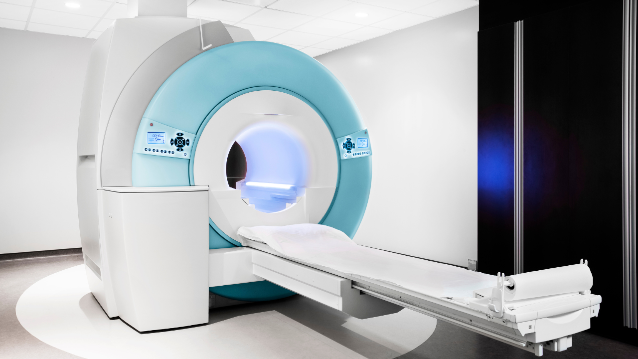
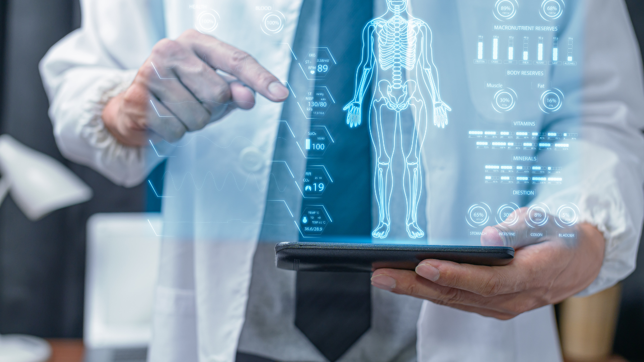
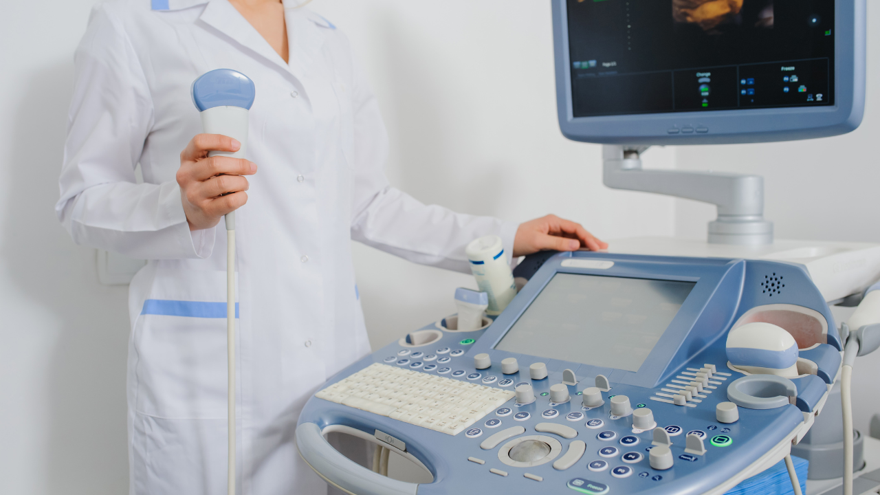
You can significantly boost diagnostic precision using advanced MRI image analysis tools. These tools enable precise image processing, integrate machine learning models, and offer real-time collaboration features. They aid in accurately identifying anatomical landmarks and differentiating between tissues, using convolutional neural networks and deep learning models. Automated algorithms detect abnormalities, isolating subtle changes in tissue morphology with high precision. Radiologists' expertise combined with these technologies ensures high-quality, patient-centered care, enhancing the accuracy and reliability of diagnoses. Exploring the full capabilities of these tools will reveal their profound impact on medical diagnostics.
Listen to the Article
Key Takeaways
- Enhanced Anomaly Detection: Machine learning algorithms improve the identification of abnormalities in MRI scans, leading to early and accurate diagnoses.
- Automated Tissue Segmentation: Advanced tools automate tissue segmentation, differentiating between structures like gray matter and white matter for precise analysis.
- Real-time Collaboration: Software with real-time collaboration features allows specialists to share insights instantly, enhancing diagnostic precision.
- Longitudinal Analysis: Integrating historical data with current findings provides a comprehensive view, aiding in tracking disease progression and treatment efficacy.
- High-Quality Visualization: Advanced image processing techniques clarify signal intensity variations, enabling detailed visualization of subtle tissue differences.
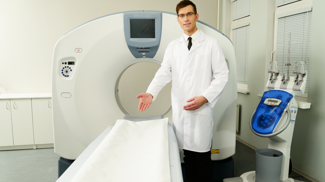
Loading Images on Workstation
Loading images onto the workstation is the crucial first step in MRI image analysis. This process requires precise calibration and compatibility checks to ensure data integrity.
You'll need to begin by verifying the MRI scanner's output format matches the workstation's accepted input formats. Ensure that the DICOM files, which are standard in medical imaging, are intact and free from corruption.
Carefully calibrate the workstation's display settings to match the MRI's output. This involves adjusting brightness and contrast for accurate representation.
Finally, confirm that all patient data is correctly anonymized to protect privacy. Skipping these steps could lead to misinterpretation of the images, impacting diagnostic decisions.
Utilizing Software Tools
To maximize the potential of MRI image analysis, you must leverage advanced software tools designed for precise image processing and interpretation. These tools, like 3D slicers and automated segmentation algorithms, allow you to scrutinize complex anatomical structures with unparalleled accuracy.
Integrating machine learning models can enhance diagnostic capabilities by recognizing subtle patterns indicative of various pathologies. You should utilize software that offers real-time collaboration features, enabling multidisciplinary teams to review findings simultaneously, thus fostering comprehensive patient care.
Additionally, customizable workflows and intuitive user interfaces streamline the analysis process, reducing the risk of human error. By employing these sophisticated tools, you ensure that every scan is meticulously examined, leading to more accurate diagnoses and ultimately improving patient outcomes.
Identifying Anatomical Landmarks
When identifying anatomical landmarks, you'll rely on advanced landmark detection algorithms to pinpoint critical structures with high precision.
Utilizing 3D visualization techniques enhances your ability to interpret spatial relationships and improves diagnostic accuracy.
Ensuring accuracy in landmark localization is crucial for effective treatment planning and follow-up assessments.
Landmark Detection Algorithms
Modern landmark detection algorithms in MRI image analysis leverage advanced machine learning techniques to accurately identify and annotate key anatomical structures.
You can utilize these algorithms to enhance diagnostic precision by automating the identification of critical landmarks such as the hippocampus, ventricles, and spinal cord.
By employing convolutional neural networks (CNNs) and deep learning models, these systems can learn from vast datasets, improving their accuracy over time.
Additionally, integrating these algorithms into clinical workflows minimizes human error and speeds up the diagnostic process.
Consequently, you'll be able to offer patients quicker and more reliable diagnoses, ensuring timely and effective treatment plans.
Embracing these innovations not only elevates the quality of care but also underscores your commitment to serving others with cutting-edge medical technology.
3D Visualization Techniques
Three-dimensional visualization techniques revolutionize how you identify anatomical landmarks in MRI images, enabling more accurate and comprehensive analysis. By rendering structures in 3D, you can explore different planes and angles, revealing hidden details and spatial relationships that 2D images might obscure.
Advanced visualization tools allow you to manipulate these images interactively, enhancing your ability to pinpoint critical landmarks. You can segment tissues, highlight specific structures, and even simulate surgical procedures. This empowerment extends to improving diagnostic precision, facilitating better patient outcomes.
Moreover, integrating these tools with machine learning algorithms further refines landmark identification, ensuring you're equipped with the most detailed and actionable insights. Ultimately, these techniques elevate the standard of care you can provide, fostering a more precise and effective diagnostic process.
Accuracy in Landmark Localization
Achieving high accuracy in landmark localization within MRI images hinges on leveraging advanced image processing algorithms and precise annotation techniques. You'll need to employ machine learning models that can learn from vast datasets to identify anatomical landmarks with pinpoint precision. Using convolutional neural networks (CNNs) enhances the ability to detect subtle variations in tissue structures.
Additionally, integrating multimodal imaging data can improve the robustness of landmark detection. Precision in annotation is crucial; manual or semi-automated methods must be meticulously validated to ensure consistency. This accuracy directly impacts diagnostic precision, enabling more effective treatment planning and patient outcomes.
Differentiating Between Tissues
MRI image analysis tools enable precise differentiation between various tissue types by exploiting differences in their magnetic properties and relaxation times. You can distinguish between gray matter, white matter, and cerebrospinal fluid by analyzing the T1 and T2 relaxation times. These tools assess the signal intensity variations, allowing you to create detailed, contrast-enhanced images. By doing so, you ensure a clearer visualization of anatomical structures.
This precision is vital for identifying subtle tissue changes that indicate various pathologies. Employing advanced algorithms and machine learning, you can automate and enhance tissue segmentation, reducing human error. Consequently, you'll improve diagnostic accuracy and patient outcomes, making your role in patient care more effective and impactful.
Detecting Abnormalities
When detecting abnormalities, you leverage advanced MRI image analysis tools to meticulously evaluate deviations in tissue structure and signal intensity that may indicate pathological conditions. These tools enable you to identify subtle changes in tissue morphology, such as the presence of lesions, tumors, or inflammation, with high precision.
You can employ segmentation algorithms to isolate suspicious regions and quantify their characteristics, including volume, shape, and texture. By comparing these parameters against established norms, you can discern abnormal tissue from healthy tissue.
Additionally, machine learning models enhance your ability to detect patterns and anomalies that might be overlooked manually. This precise detection process allows you to provide early and accurate diagnoses, ultimately contributing to better patient outcomes and more effective treatments.
Annotating MRI Images
Annotating MRI images involves meticulously labeling anatomical structures and pathological findings to facilitate accurate interpretation and analysis. You need to ensure that each label is accurate, as this precision directly impacts patient care.
The process includes:
- Identifying key anatomical landmarks: Properly marking features like ventricles, lobes, and vessels.
- Highlighting abnormalities: Pointing out lesions, tumors, or other pathological entities.
- Using standardized nomenclature: Ensuring consistency in labels to avoid misinterpretation.
- Employing advanced software tools: Utilizing AI and machine learning for more efficient annotation.
- Collaborating with specialists: Working closely with radiologists and other experts to ensure accuracy.
Comparing With Prior Scans
To gain a comprehensive understanding of a patient's condition, it's crucial to meticulously compare current MRI scans with prior images, noting any changes in anatomical structures or pathological findings.
By employing advanced image analysis tools, you can detect subtle variations that might signify disease progression or treatment response.
Automated algorithms can highlight differences in tissue density, lesion size, and other clinically relevant metrics, enabling quicker and more accurate assessments.
This practice not only refines diagnostic precision but also helps in tracking the efficacy of therapeutic interventions.
By integrating historical data with present findings, you ensure a longitudinal perspective, facilitating personalized care plans that are responsive to the patient's evolving medical needs.
This approach embodies a commitment to high-quality, patient-centered healthcare.
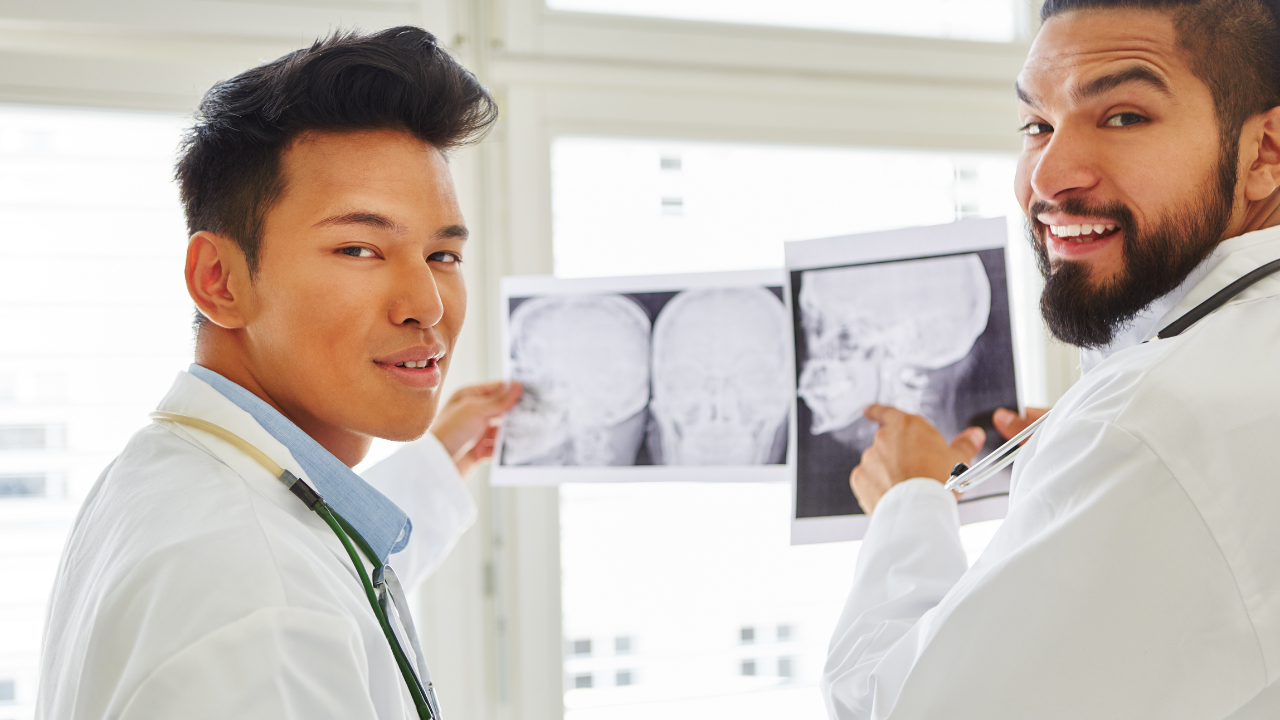
Consulting With Radiologists
Collaborating with radiologists enhances the diagnostic process by leveraging their specialized expertise in interpreting complex MRI images. You can significantly improve diagnostic accuracy and patient outcomes by consulting with these experts. Radiologists provide insights that are crucial for understanding intricate anatomical details and identifying subtle abnormalities.
- Expert Interpretation: Radiologists' training allows for precise analysis of MRI images, reducing diagnostic errors.
- Clinical Correlation: They correlate MRI findings with clinical data, offering a comprehensive diagnostic overview.
- Technological Proficiency: Radiologists are adept at utilizing advanced MRI analysis tools, optimizing their use.
- Anomaly Detection: Their experience enables the identification of rare and complex pathologies missed by automated systems.
- Continual Learning: They stay updated with the latest advancements in MRI technology, ensuring cutting-edge diagnostic practices.
This collaboration ultimately serves the best interests of your patients.
Generating and Archiving Reports
Generating and archiving reports in MRI image analysis entails creating detailed, accurate documentation of findings and securely storing these records for future reference and clinical use. You must ensure that each report captures essential diagnostic information, including observed abnormalities, measurements, and interpretative insights.
Advanced software tools can automate parts of this process, reducing human error and enhancing consistency. Archiving involves using secure, compliant digital storage solutions to maintain accessibility and confidentiality. Properly archived reports support longitudinal studies, assist in patient follow-ups, and facilitate collaborative care.
Recent Posts
Office Hours:
Monday-Friday
8:00am-5:00pm
Saturday by appointment
Services
Contact Details
Address: 1971 Gowdey Road,
Naperville, IL 60563
Phone: 630-416-1300
Fax:
630-416-1511
Email: info@foxvalleyimaging.com
© Copyright 2023 Fox Valley Imaging, Inc..